 Movie
Movie Controller

[English] 日本語
 Yorodumi
- PDB-5ft1: Crystal structure of gp37(Dip) from bacteriophage phiKZ bound to ... -
+ Open data

- Basic information
| Entry | Database: PDB / ID: 5ft1 | ||||||
|---|---|---|---|---|---|---|---|
| Title | Crystal structure of gp37(Dip) from bacteriophage phiKZ bound to RNase E of Pseudomonas aeruginosa | ||||||
 Components Components |
| ||||||
 Keywords Keywords | HYDROLASE/INHIBITOR / HYDROLASE-INHIBITOR COMPLEX / DIP / PHIKZ /  BACTERIOPHAGE / RNASE E / RIBONUCLEASE INHIBITOR / RNA DEGRADOSOME / PSEUDOMONAS AERUGINOSA BACTERIOPHAGE / RNASE E / RIBONUCLEASE INHIBITOR / RNA DEGRADOSOME / PSEUDOMONAS AERUGINOSA | ||||||
| Function / homology |  Function and homology informationribonuclease E / ribonuclease E activity / tRNA processing / mRNA catabolic process / RNA nuclease activity / RNA endonuclease activity / cytoplasmic side of plasma membrane / rRNA processing / tRNA binding / rRNA binding ...ribonuclease E / ribonuclease E activity / tRNA processing / mRNA catabolic process / RNA nuclease activity / RNA endonuclease activity / cytoplasmic side of plasma membrane / rRNA processing / tRNA binding / rRNA binding / magnesium ion binding / zinc ion binding / cytoplasm Function and homology informationribonuclease E / ribonuclease E activity / tRNA processing / mRNA catabolic process / RNA nuclease activity / RNA endonuclease activity / cytoplasmic side of plasma membrane / rRNA processing / tRNA binding / rRNA binding ...ribonuclease E / ribonuclease E activity / tRNA processing / mRNA catabolic process / RNA nuclease activity / RNA endonuclease activity / cytoplasmic side of plasma membrane / rRNA processing / tRNA binding / rRNA binding / magnesium ion binding / zinc ion binding / cytoplasmSimilarity search - Function | ||||||
| Biological species |  PSEUDOMONAS PHAGE PHIKZ (virus) PSEUDOMONAS PHAGE PHIKZ (virus)  PSEUDOMONAS AERUGINOSA (bacteria) PSEUDOMONAS AERUGINOSA (bacteria) | ||||||
| Method | X-RAY DIFFRACTION / SYNCHROTRON / MOLECULAR REPLACEMENT / Resolution: 2.75 Å | ||||||
 Authors Authors | Van den Bossche, A. / Hardwick, S.W. / Ceyssens, P.J. / Hendrix, H. / Voet, M. / Dendooven, T. / Bandyra, K.J. / De Maeyer, M. / Aertsen, A. / Noben, J.P. ...Van den Bossche, A. / Hardwick, S.W. / Ceyssens, P.J. / Hendrix, H. / Voet, M. / Dendooven, T. / Bandyra, K.J. / De Maeyer, M. / Aertsen, A. / Noben, J.P. / Luisi, B.F. / Lavigne, R. | ||||||
 Citation Citation | Journal: Elife / Year: 2016 Title: Structural elucidation of a novel mechanism for the bacteriophage-based inhibition of the RNA degradosome. Authors: Van den Bossche, A. / Hardwick, S.W. / Ceyssens, P.J. / Hendrix, H. / Voet, M. / Dendooven, T. / Bandyra, K.J. / De Maeyer, M. / Aertsen, A. / Noben, J.P. / Luisi, B.F. / Lavigne, R. | ||||||
| History |
|
- Structure visualization
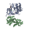
| Structure viewer | Molecule: MolmilJmol/JSmol |
|---|
- Downloads & links
-Download
| PDBx/mmCIF format | 5ft1.cif.gz | 298.8 KB | Display | PDBx/mmCIF format |
|---|---|---|---|---|
| PDB format | pdb5ft1.ent.gz | 239.6 KB | Display | PDB format |
| PDBx/mmJSON format | 5ft1.json.gz | Tree view | PDBx/mmJSON format | |
| Others |  Other downloads Other downloads |
-Validation report
| Arichive directory | https://data.pdbj.org/pub/pdb/validation_reports/ft/5ft1ftp://data.pdbj.org/pub/pdb/validation_reports/ft/5ft1 | HTTPS FTP |
|---|
-Related structure data
| Related structure data |  5ft0SC S: Starting model for refinement C: citing same article ( |
|---|---|
| Similar structure data |


-Links
 PDBj
PDBj


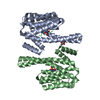
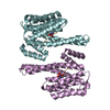
- Assembly
| Deposited unit | 
| ||||||||
|---|---|---|---|---|---|---|---|---|---|
| 1 | 
| ||||||||
| 2 | 
| ||||||||
| 3 | 
| ||||||||
| Unit cell |
|
-Components
| #1: Protein | Mass: 31991.303 Da / Num. of mol.: 6 Source method: isolated from a genetically manipulated source Source: (gene. exp.) PSEUDOMONAS PHAGE PHIKZ (virus) / Production host: ESCHERICHIA COLI (E. coli) / Strain (production host): BL21(DE3) / Variant (production host): PLYSS / References: UniProt: Q8SDC5#2: Protein/peptide | / RNASE EMass: 2704.057 Da / Num. of mol.: 6 / Fragment: RESIDUES 756-775 / Source method: obtained synthetically / Source: (synth.) PSEUDOMONAS AERUGINOSA (bacteria) / References: UniProt: Q9HZM8, ribonuclease E#3: Water | ChemComp-HOH / | Water Mass: 18.015 Da / Num. of mol.: 1 / Source method: isolated from a natural source / Formula: H2O Mass: 18.015 Da / Num. of mol.: 1 / Source method: isolated from a natural source / Formula: H2O |
|---|
-Experimental details
-Experiment
| Experiment | Method: X-RAY DIFFRACTION / Number of used crystals: 1 |
|---|
- Sample preparation
| Crystal | Density Matthews: 2.37 Å3/Da / Density % sol: 48.16 % Description: DATA PROCESSED IN LOW SYMMETRY P1 SPACE GROUP AS REFINEMENT STATISTICS AND ELECTRON DENSITY FOR BOUND PEPTIDE WERE MARGINALLY BETTER THAN IN RECOMMENDED H32 SPACE GROUP |
|---|---|
| Crystal grow | Details: 100 MM KH2PO4, 100 MM NAH2PO4, 100 MM MES PH 6.0, 300 MM NACL, 0.2 M SODIUM THIOCYANATE |
-Data collection
| Diffraction | Mean temperature: 100 K |
|---|---|
| Diffraction source | Source: SYNCHROTRON / Site: Diamond  / Beamline: I04 / Wavelength: 0.9793 / Beamline: I04 / Wavelength: 0.9793 |
| Detector | Type: DECTRIS PILATUS 6M / Detector: PIXEL / Date: Jul 1, 2015 |
| Radiation | Protocol: SINGLE WAVELENGTH / Monochromatic (M) / Laue (L): M / Scattering type: x-ray |
| Radiation wavelength | Wavelength: 0.9793 Å / Relative weight: 1 |
| Reflection twin | Operator: -L,-K,-H / Fraction: 0.5 |
| Reflection | Resolution: 2.75→72.2 Å / Num. obs: 39553 / % possible obs: 94.2 % / Observed criterion σ(I): 2 / Redundancy: 1.9 % / Rmerge(I) obs: 0.03 / Net I/σ(I): 12.3 |
| Reflection shell | Resolution: 2.75→2.84 Å / Redundancy: 1.8 % / Rmerge(I) obs: 0.43 / Mean I/σ(I) obs: 1.7 / % possible all: 93.2 |
- Processing
| Software |
| ||||||||||||||||||||||||||||||||||||||||||||||||||||||||||||||||||||||||||||||||||||||||||||||||||||||||||||||||
|---|---|---|---|---|---|---|---|---|---|---|---|---|---|---|---|---|---|---|---|---|---|---|---|---|---|---|---|---|---|---|---|---|---|---|---|---|---|---|---|---|---|---|---|---|---|---|---|---|---|---|---|---|---|---|---|---|---|---|---|---|---|---|---|---|---|---|---|---|---|---|---|---|---|---|---|---|---|---|---|---|---|---|---|---|---|---|---|---|---|---|---|---|---|---|---|---|---|---|---|---|---|---|---|---|---|---|---|---|---|---|---|---|---|
| Refinement | Method to determine structure: MOLECULAR REPLACEMENT Starting model: PDB ENTRY 5FT0 Resolution: 2.75→72.188 Å / σ(F): 1.98 / Phase error: 32.82 / Stereochemistry target values: TWIN_LSQ_F
| ||||||||||||||||||||||||||||||||||||||||||||||||||||||||||||||||||||||||||||||||||||||||||||||||||||||||||||||||
| Solvent computation | Shrinkage radii: 0.9 Å / VDW probe radii: 1.11 Å / Solvent model: FLAT BULK SOLVENT MODEL | ||||||||||||||||||||||||||||||||||||||||||||||||||||||||||||||||||||||||||||||||||||||||||||||||||||||||||||||||
| Refinement step | Cycle: LAST / Resolution: 2.75→72.188 Å
| ||||||||||||||||||||||||||||||||||||||||||||||||||||||||||||||||||||||||||||||||||||||||||||||||||||||||||||||||
| Refine LS restraints |
| ||||||||||||||||||||||||||||||||||||||||||||||||||||||||||||||||||||||||||||||||||||||||||||||||||||||||||||||||
| LS refinement shell |
|
