 Movie
Movie Controller

[English] 日本語
 Yorodumi
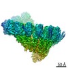
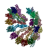
- PDB-5fm1: Structure of gamma-tubulin small complex based on a cryo-EM map, ... -
+ Open data

- Basic information
| Entry | Database: PDB / ID: 5fm1 | ||||||
|---|---|---|---|---|---|---|---|
| Title | Structure of gamma-tubulin small complex based on a cryo-EM map, chemical cross-links, and a remotely related structure | ||||||
 Components Components |
| ||||||
 Keywords Keywords | CELL CYCLE / MICROTUBULE / NUCLEATION / TUBULIN / FILAMENT | ||||||
| Function / homology |  Function and homology information Function and homology informationinner plaque of spindle pole body / microtubule nucleation by spindle pole body / outer plaque of spindle pole body / gamma-tubulin small complex / regulation of microtubule nucleation / equatorial microtubule organizing center / mitotic spindle pole body / gamma-tubulin complex / meiotic spindle organization / microtubule nucleation ...inner plaque of spindle pole body / microtubule nucleation by spindle pole body / outer plaque of spindle pole body / gamma-tubulin small complex / regulation of microtubule nucleation / equatorial microtubule organizing center / mitotic spindle pole body / gamma-tubulin complex / meiotic spindle organization / microtubule nucleation / positive regulation of cytoplasmic translation / gamma-tubulin binding / spindle pole body / mitotic sister chromatid segregation / spindle assembly / meiotic cell cycle / cytoplasmic microtubule organization / mitotic spindle organization / structural constituent of cytoskeleton / spindle pole / spindle / mitotic cell cycle / microtubule / GTP binding / nucleus / cytoplasm Similarity search - Function | ||||||
| Biological species |  | ||||||
| Method | ELECTRON MICROSCOPY / helical reconstruction / cryo EM / Resolution: 8 Å | ||||||
 Authors Authors | Greenberg, C.H. / Kollman, J. / Zelter, A. / Johnson, R. / MacCoss, M.J. / Davis, T.N. / Agard, D.A. / Sali, A. | ||||||
 Citation Citation | Journal: J Struct Biol / Year: 2016 Title: Structure of γ-tubulin small complex based on a cryo-EM map, chemical cross-links, and a remotely related structure. Authors: Charles H Greenberg / Justin Kollman / Alex Zelter / Richard Johnson / Michael J MacCoss / Trisha N Davis / David A Agard / Andrej Sali /  Abstract: Modeling protein complex structures based on distantly related homologues can be challenging due to poor sequence and structure conservation. Therefore, utilizing even low-resolution experimental ...Modeling protein complex structures based on distantly related homologues can be challenging due to poor sequence and structure conservation. Therefore, utilizing even low-resolution experimental data can significantly increase model precision and accuracy. Here, we present models of the two key functional states of the yeast γ-tubulin small complex (γTuSC): one for the low-activity "open" state and another for the higher-activity "closed" state. Both models were computed based on remotely related template structures and cryo-EM density maps at 6.9Å and 8.0Å resolution, respectively. For each state, extensive sampling of alignments and conformations was guided by the fit to the corresponding cryo-EM density map. The resulting good-scoring models formed a tightly clustered ensemble of conformations in most regions. We found significant structural differences between the two states, primarily in the γ-tubulin subunit regions where the microtubule binds. We also report a set of chemical cross-links that were found to be consistent with equilibrium between the open and closed states. The protocols developed here have been incorporated into our open-source Integrative Modeling Platform (IMP) software package (http://integrativemodeling.org), and can therefore be applied to many other systems. #1: Journal: Nature / Year: 2010Title: Microtubule nucleating gamma-TuSC assembles structures with 13-fold microtubule-like symmetry. Authors: Justin M Kollman / Jessica K Polka / Alex Zelter / Trisha N Davis / David A Agard / Abstract: Microtubules are nucleated in vivo by gamma-tubulin complexes. The 300-kDa gamma-tubulin small complex (gamma-TuSC), consisting of two molecules of gamma-tubulin and one copy each of the accessory ...Microtubules are nucleated in vivo by gamma-tubulin complexes. The 300-kDa gamma-tubulin small complex (gamma-TuSC), consisting of two molecules of gamma-tubulin and one copy each of the accessory proteins Spc97 and Spc98, is the conserved, essential core of the microtubule nucleating machinery. In metazoa multiple gamma-TuSCs assemble with other proteins into gamma-tubulin ring complexes (gamma-TuRCs). The structure of gamma-TuRC indicated that it functions as a microtubule template. Because each gamma-TuSC contains two molecules of gamma-tubulin, it was assumed that the gamma-TuRC-specific proteins are required to organize gamma-TuSCs to match 13-fold microtubule symmetry. Here we show that Saccharomyces cerevisiae gamma-TuSC forms rings even in the absence of other gamma-TuRC components. The yeast adaptor protein Spc110 stabilizes the rings into extended filaments and is required for oligomer formation under physiological buffer conditions. The 8-A cryo-electron microscopic reconstruction of the filament reveals 13 gamma-tubulins per turn, matching microtubule symmetry, with plus ends exposed for interaction with microtubules, implying that one turn of the filament constitutes a microtubule template. The domain structures of Spc97 and Spc98 suggest functions for conserved sequence motifs, with implications for the gamma-TuRC-specific proteins. The gamma-TuSC filaments nucleate microtubules at a low level, and the structure provides a strong hypothesis for how nucleation is regulated, converting this less active form to a potent nucleator. | ||||||
| History |
|
- Structure visualization
| Movie |
Movie viewer |
|---|---|
| Structure viewer | Molecule: MolmilJmol/JSmol |
- Downloads & links
-Download
| PDBx/mmCIF format | 5fm1.cif.gz | 3.3 MB | Display | PDBx/mmCIF format |
|---|---|---|---|---|
| PDB format | pdb5fm1.ent.gz | 2.8 MB | Display | PDB format |
| PDBx/mmJSON format | 5fm1.json.gz | Tree view | PDBx/mmJSON format | |
| Others |  Other downloads Other downloads |
-Validation report
| Summary document | 5fm1_validation.pdf.gz | 1.2 MB | Display | wwPDB validaton report |
|---|---|---|---|---|
| Full document | 5fm1_full_validation.pdf.gz | 2.2 MB | Display | |
| Data in XML | 5fm1_validation.xml.gz | 490.6 KB | Display | |
| Data in CIF | 5fm1_validation.cif.gz | 724.8 KB | Display | |
| Arichive directory | https://data.pdbj.org/pub/pdb/validation_reports/fm/5fm1ftp://data.pdbj.org/pub/pdb/validation_reports/fm/5fm1 | HTTPS FTP |
-Related structure data
| Related structure data | 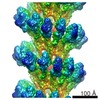 1731M  5flzC C: citing same article ( M: map data used to model this data |
|---|---|
| Similar structure data |

-Links
 PDBj
PDBj


- Assembly
| Deposited unit | 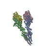
|
|---|---|
| 1 | x 7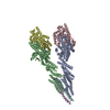
|
| Number of models | 10 |
-Components
| #1: Protein | Mass: 96940.594 Da / Num. of mol.: 1 Source method: isolated from a genetically manipulated source Source: (gene. exp.) Plasmid: PDV45 / Production host:   SPODOPTERA FRUGIPERDA (fall armyworm) / References: UniProt: P38863 SPODOPTERA FRUGIPERDA (fall armyworm) / References: UniProt: P38863 | ||||
|---|---|---|---|---|---|
| #2: Protein | Mass: 98336.211 Da / Num. of mol.: 1 Source method: isolated from a genetically manipulated source Source: (gene. exp.) Plasmid: PDV45 / Production host: SPODOPTERA FRUGIPERDA (fall armyworm) / References: UniProt: P53540 | ||||
| #3: Protein | Mass: 52671.188 Da / Num. of mol.: 2 Source method: isolated from a genetically manipulated source Source: (gene. exp.) Plasmid: PDV45 / Production host: SPODOPTERA FRUGIPERDA (fall armyworm) / References: UniProt: P53378#4: Protein/peptide | Mass: 3762.629 Da / Num. of mol.: 2 Source method: isolated from a genetically manipulated source Details: CONSTRUCT INCLUDES THE FIRST 220 RESIDUES OF SPC110P IT CANNOT BE UNIQUELY IDENTIFIED WHICH FRAGMENT OF SPC110 IS PRESENT IN THE MAP, THUS A POLY-ALANINE HELIX WAS FITTED. Source: (gene. exp.) Plasmid: PDV45 / Production host: SPODOPTERA FRUGIPERDA (fall armyworm)Sequence details | ONLY RESIDUES 1-220 INCLUDED IN STRUCTURE. CHAINS E AND F CORRESPOND | |
-Experimental details
-Experiment
| Experiment | Method: ELECTRON MICROSCOPY |
|---|---|
| EM experiment | Aggregation state: FILAMENT / 3D reconstruction method: helical reconstruction |
- Sample preparation
| Component | Name: GAMMA TUBULIN SMALL COMPLEX / Type: COMPLEX |
|---|---|
| Buffer solution | Name: 100 MM KCL, 1MM GTP, 1MM MGCL2, 2MM EGTA, 40 MM HEPES / pH: 7.6 Details: 100 MM KCL, 1MM GTP, 1MM MGCL2, 2MM EGTA, 40 MM HEPES |
| Specimen | Conc.: 2 mg/ml / Embedding applied: NO / Shadowing applied: NO / Staining applied: NO / Vitrification applied: YES |
| Specimen support | Details: OTHER |
| Vitrification | Instrument: FEI VITROBOT MARK I / Cryogen name: ETHANE / Details: LIQUID ETHANE |
- Electron microscopy imaging
| Experimental equipment |  Model: Tecnai F20 / Image courtesy: FEI Company |
|---|---|
| Microscopy | Model: FEI TECNAI F20 / Date: Sep 23, 2009 |
| Electron gun | Electron source:  FIELD EMISSION GUN / Accelerating voltage: 200 kV / Illumination mode: FLOOD BEAM FIELD EMISSION GUN / Accelerating voltage: 200 kV / Illumination mode: FLOOD BEAM |
| Electron lens | Mode: BRIGHT FIELD / Nominal magnification: 60000 X / Nominal defocus max: 3000 nm / Nominal defocus min: 800 nm / Cs: 2.2 mm |
| Image recording | Electron dose: 20 e/Å2 / Film or detector model: TVIPS TEMCAM-F816 (8k x 8k) |
- Processing
| EM software |
| |||||||||||||||
|---|---|---|---|---|---|---|---|---|---|---|---|---|---|---|---|---|
| CTF correction | Details: WHOLE MICROGRAPH | |||||||||||||||
| 3D reconstruction | Method: ITERATIVE HELICAL REAL SPACE RECONSTRUCTION / Resolution: 8 Å Details: INITIAL MODELS WERE GENERATED WITH MODELER 9.15 BASED ON PDB ENTRIES 3RIP (FOR GCP2 AND GCP3) AND 3CB2 (FOR GAMMA TUBULIN). TWO COMPLETE GAMMA-TUSC STRUCTURES WERE MODELED SIMULTANEOUSLY, ...Details: INITIAL MODELS WERE GENERATED WITH MODELER 9.15 BASED ON PDB ENTRIES 3RIP (FOR GCP2 AND GCP3) AND 3CB2 (FOR GAMMA TUBULIN). TWO COMPLETE GAMMA-TUSC STRUCTURES WERE MODELED SIMULTANEOUSLY, RIGIDLY FITTED INTO THE CRYO-EM MAP WITH UCSF CHIMERA, THEN FLEXIBLY FITTED WITH MDFF. ADDITIONAL SECONDARY STRUCTURE AND SYMMETRY RESTRAINTS WERE ADDED. FINAL VALIDATION PERFORMED WITH THE INTEGRATIVE MODELING PLATFORM. FOR DETAILS, ALL CODE IS DEPOSITED AT GITHUB. COMSLASHINTEGRATIVEMODELINGSLASHGAMMA-TUSC SUBMISSION BASED ON EXPERIMENTAL DATA FROM EMDB EMD-1731. (DEPOSITION ID: 7273). Symmetry type: HELICAL | |||||||||||||||
| Atomic model building | Protocol: FLEXIBLE FIT / Space: REAL Details: METHOD--FLEXIBLE FITTING REFINEMENT PROTOCOL--X-RAY | |||||||||||||||
| Refinement | Highest resolution: 8 Å | |||||||||||||||
| Refinement step | Cycle: LAST / Highest resolution: 8 Å
|
