 Movie
Movie Controller

[English] 日本語
 Yorodumi
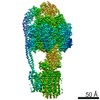
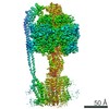
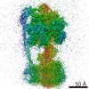
- PDB-5fl7: Structure of the F1c10 complex from Yarrowia lipolytica ATP synthase -
+ Open data

- Basic information
| Entry | Database: PDB / ID: 5fl7 | ||||||
|---|---|---|---|---|---|---|---|
| Title | Structure of the F1c10 complex from Yarrowia lipolytica ATP synthase | ||||||
 Components Components | (ATP SYNTHASE ...) x 6 | ||||||
 Keywords Keywords | HYDROLASE / ATP SYNTHASE FAMILY / NUCLEOTIDE BINDING / PROTON TRANSPORTING / ROTATIONAL MECHANISM / ATP BIOSYNTHETIC PROCESS / ATP SYNTHESIS/HYDROLYSIS | ||||||
| Function / homology |  Function and homology information Function and homology information: / : / proton-transporting ATP synthase complex, coupling factor F(o) / : / proton transmembrane transporter activity / proton motive force-driven ATP synthesis / proton-transporting ATP synthase complex, catalytic core F(1) / H+-transporting two-sector ATPase / proton-transporting ATPase activity, rotational mechanism / proton-transporting ATP synthase activity, rotational mechanism ...: / : / proton-transporting ATP synthase complex, coupling factor F(o) / : / proton transmembrane transporter activity / proton motive force-driven ATP synthesis / proton-transporting ATP synthase complex, catalytic core F(1) / H+-transporting two-sector ATPase / proton-transporting ATPase activity, rotational mechanism / proton-transporting ATP synthase activity, rotational mechanism / proton motive force-driven mitochondrial ATP synthesis / ADP binding / membrane => GO:0016020 / mitochondrial inner membrane / lipid binding / ATP binding / plasma membrane Similarity search - Function | ||||||
| Biological species |  YARROWIA LIPOLYTICA (yeast) YARROWIA LIPOLYTICA (yeast) | ||||||
| Method |  X-RAY DIFFRACTION / SYNCHROTRON / MOLECULAR REPLACEMENT / Resolution: 3.5 Å X-RAY DIFFRACTION / SYNCHROTRON / MOLECULAR REPLACEMENT / Resolution: 3.5 Å | ||||||
 Authors Authors | Parey, K. / Bublitz, M. / Meier, T. | ||||||
 Citation Citation | Journal: Mol Cell / Year: 2016 Title: Structure of a Complete ATP Synthase Dimer Reveals the Molecular Basis of Inner Mitochondrial Membrane Morphology. Authors: Alexander Hahn / Kristian Parey / Maike Bublitz / Deryck J Mills / Volker Zickermann / Janet Vonck / Werner Kühlbrandt / Thomas Meier /  Abstract: We determined the structure of a complete, dimeric F1Fo-ATP synthase from yeast Yarrowia lipolytica mitochondria by a combination of cryo-EM and X-ray crystallography. The final structure resolves 58 ...We determined the structure of a complete, dimeric F1Fo-ATP synthase from yeast Yarrowia lipolytica mitochondria by a combination of cryo-EM and X-ray crystallography. The final structure resolves 58 of the 60 dimer subunits. Horizontal helices of subunit a in Fo wrap around the c-ring rotor, and a total of six vertical helices assigned to subunits a, b, f, i, and 8 span the membrane. Subunit 8 (A6L in human) is an evolutionary derivative of the bacterial b subunit. On the lumenal membrane surface, subunit f establishes direct contact between the two monomers. Comparison with a cryo-EM map of the F1Fo monomer identifies subunits e and g at the lateral dimer interface. They do not form dimer contacts but enable dimer formation by inducing a strong membrane curvature of ∼100°. Our structure explains the structural basis of cristae formation in mitochondria, a landmark signature of eukaryotic cell morphology. | ||||||
| History |
| ||||||
| Remark 700 | SHEET DETERMINATION METHOD: DSSP THE SHEETS PRESENTED AS "AA" IN EACH CHAIN ON SHEET RECORDS BELOW ... SHEET DETERMINATION METHOD: DSSP THE SHEETS PRESENTED AS "AA" IN EACH CHAIN ON SHEET RECORDS BELOW IS ACTUALLY AN 6-STRANDED BARREL THIS IS REPRESENTED BY A 7-STRANDED SHEET IN WHICH THE FIRST AND LAST STRANDS ARE IDENTICAL. THE SHEETS PRESENTED AS "BA" IN EACH CHAIN ON SHEET RECORDS BELOW IS ACTUALLY AN 10-STRANDED BARREL THIS IS REPRESENTED BY A 11-STRANDED SHEET IN WHICH THE FIRST AND LAST STRANDS ARE IDENTICAL. THE SHEETS PRESENTED AS "DA" IN EACH CHAIN ON SHEET RECORDS BELOW IS ACTUALLY AN 6-STRANDED BARREL THIS IS REPRESENTED BY A 7-STRANDED SHEET IN WHICH THE FIRST AND LAST STRANDS ARE IDENTICAL. THE SHEETS PRESENTED AS "EA" IN EACH CHAIN ON SHEET RECORDS BELOW IS ACTUALLY AN 6-STRANDED BARREL THIS IS REPRESENTED BY A 7-STRANDED SHEET IN WHICH THE FIRST AND LAST STRANDS ARE IDENTICAL. |
- Structure visualization
| Structure viewer | Molecule: MolmilJmol/JSmol |
|---|
- Downloads & links
-Download
| PDBx/mmCIF format | 5fl7.cif.gz | 748.3 KB | Display | PDBx/mmCIF format |
|---|---|---|---|---|
| PDB format | pdb5fl7.ent.gz | 609.9 KB | Display | PDB format |
| PDBx/mmJSON format | 5fl7.json.gz | Tree view | PDBx/mmJSON format | |
| Others |  Other downloads Other downloads |
-Validation report
| Summary document | 5fl7_validation.pdf.gz | 1.6 MB | Display | wwPDB validaton report |
|---|---|---|---|---|
| Full document | 5fl7_full_validation.pdf.gz | 1.7 MB | Display | |
| Data in XML | 5fl7_validation.xml.gz | 131.4 KB | Display | |
| Data in CIF | 5fl7_validation.cif.gz | 177.3 KB | Display | |
| Arichive directory | https://data.pdbj.org/pub/pdb/validation_reports/fl/5fl7ftp://data.pdbj.org/pub/pdb/validation_reports/fl/5fl7 | HTTPS FTP |
-Related structure data
| Related structure data | 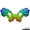 8151C 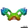 8152C 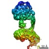 8153C 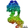 8154C  8155C  2xokS S: Starting model for refinement C: citing same article ( |
|---|---|
| Similar structure data |


-Links
 PDBj
PDBj


- Assembly
| Deposited unit | 
| ||||||||
|---|---|---|---|---|---|---|---|---|---|
| 1 |
| ||||||||
| Unit cell |
|
-Components
-ATP SYNTHASE ... , 6 types, 19 molecules ABCDEFGHIKLMNOPQRST
| #1: Protein | Mass: 58147.430 Da / Num. of mol.: 3 / Source method: isolated from a natural source / Source: (natural) YARROWIA LIPOLYTICA (yeast) / References: UniProt: Q6C326#2: Protein | Mass: 54590.828 Da / Num. of mol.: 3 / Source method: isolated from a natural source / Source: (natural) YARROWIA LIPOLYTICA (yeast)References: UniProt: Q6CFT7, H+-transporting two-sector ATPase #3: Protein | | Mass: 32378.797 Da / Num. of mol.: 1 / Source method: isolated from a natural source / Source: (natural) YARROWIA LIPOLYTICA (yeast) / References: UniProt: Q6C338, EC: 3.6.1.34#4: Protein | | Mass: 14811.274 Da / Num. of mol.: 1 / Source method: isolated from a natural source / Source: (natural) YARROWIA LIPOLYTICA (yeast) / References: UniProt: Q6C877, EC: 3.6.1.34#5: Protein/peptide | | Mass: 1379.692 Da / Num. of mol.: 1 / Source method: isolated from a natural source / Source: (natural) YARROWIA LIPOLYTICA (yeast) / References: EC: 3.6.1.34#6: Protein | Mass: 7719.265 Da / Num. of mol.: 10 / Source method: isolated from a natural source / Source: (natural) YARROWIA LIPOLYTICA (yeast)References: UniProt: Q37695, H+-transporting two-sector ATPase |
|---|
-Non-polymers , 4 types, 27 molecules 


| #7: Chemical |  Mass: 507.181 Da / Num. of mol.: 3 / Source method: obtained synthetically / Formula: C10H16N5O13P3 / Comment: ATP, energy-carrying molecule*YM Mass: 507.181 Da / Num. of mol.: 3 / Source method: obtained synthetically / Formula: C10H16N5O13P3 / Comment: ATP, energy-carrying molecule*YM#8: Chemical | ChemComp-MG /  Mass: 24.305 Da / Num. of mol.: 5 / Source method: obtained synthetically / Formula: Mg Mass: 24.305 Da / Num. of mol.: 5 / Source method: obtained synthetically / Formula: Mg#9: Chemical |  Mass: 427.201 Da / Num. of mol.: 2 / Source method: obtained synthetically / Formula: C10H15N5O10P2 / Comment: ADP, energy-carrying molecule*YM Mass: 427.201 Da / Num. of mol.: 2 / Source method: obtained synthetically / Formula: C10H15N5O10P2 / Comment: ADP, energy-carrying molecule*YM#10: Water | ChemComp-HOH / | Mass: 18.015 Da / Num. of mol.: 17 / Source method: isolated from a natural source / Formula: H2O |
|---|
-Details
| Sequence details | CHAIN I NOT ANNOTATED IN THE UNIPROT DATABANK |
|---|
-Experimental details
-Experiment
| Experiment | Method: X-RAY DIFFRACTION / Number of used crystals: 1 |
|---|
- Sample preparation
| Crystal | Density Matthews: 2.32 Å3/Da / Density % sol: 47 % / Description: NONE |
|---|---|
| Crystal grow | pH: 8 / Details: 100 MM TRIS-HCL, POLYETHYLENE GLYCOL 400, pH 8.0 |
-Data collection
| Diffraction | Mean temperature: 100 K |
|---|---|
| Diffraction source | Source: SYNCHROTRON / Site: SLS  / Beamline: X10SA / Wavelength: 1.00774 / Beamline: X10SA / Wavelength: 1.00774 |
| Detector | Type: DECTRIS PILATUS 6M / Detector: PIXEL / Date: Mar 5, 2015 / Details: MIRRORS |
| Radiation | Monochromator: SI(111) MONOCHROMATOR / Protocol: SINGLE WAVELENGTH / Monochromatic (M) / Laue (L): M / Scattering type: x-ray |
| Radiation wavelength | Wavelength: 1.00774 Å / Relative weight: 1 |
| Reflection | Resolution: 3.5→49.19 Å / Num. obs: 75882 / % possible obs: 99.9 % / Observed criterion σ(I): -3 / Redundancy: 19.47 % / Biso Wilson estimate: 157.51 Å2 / Rmerge(I) obs: 0.18 / Net I/σ(I): 9.84 |
| Reflection shell | Resolution: 3.5→3.6 Å / Redundancy: 19.8 % / Mean I/σ(I) obs: 0.61 / % possible all: 100 |
- Processing
| Software |
| |||||||||||||||||||||||||||||||||||||||||||||||||||||||||||||||||||||||||||||||||||||||||||||||||||||||||
|---|---|---|---|---|---|---|---|---|---|---|---|---|---|---|---|---|---|---|---|---|---|---|---|---|---|---|---|---|---|---|---|---|---|---|---|---|---|---|---|---|---|---|---|---|---|---|---|---|---|---|---|---|---|---|---|---|---|---|---|---|---|---|---|---|---|---|---|---|---|---|---|---|---|---|---|---|---|---|---|---|---|---|---|---|---|---|---|---|---|---|---|---|---|---|---|---|---|---|---|---|---|---|---|---|---|---|
| Refinement | Method to determine structure: MOLECULAR REPLACEMENT Starting model: PDB ENTRY 2XOK Resolution: 3.5→49.366 Å / SU ML: 0.64 / σ(F): 1.34 / Phase error: 38.06 / Stereochemistry target values: ML Details: THE FOLLOWING RESIDUES ARE DISORDERED AND HAVE NOT BEEN MODELLED CHAIN A 1-48 534- -536 CHAIN B 1-48 429-434 534-536 CHAIN C 1-48 534-536 CHAIN D 1-36 507-510 CHAIN E 1-38 507-510 CHAIN F 1- ...Details: THE FOLLOWING RESIDUES ARE DISORDERED AND HAVE NOT BEEN MODELLED CHAIN A 1-48 534- -536 CHAIN B 1-48 429-434 534-536 CHAIN C 1-48 534-536 CHAIN D 1-36 507-510 CHAIN E 1-38 507-510 CHAIN F 1-36 507-510 CHAIN G 1-22 117-119 293 CHAIN H 1-14 95-100 134- -136 CHAIN I WAS MODELLED AND REFINED AS POLY-ALANINE CHAIN K 1 75-76 CHAIN L 1 74-76 CHAIN M 1 CHAIN N 76 CHAIN O 1 CHAIN P 76 CHAIN R 1 76 CHAIN S 1 76 CHAIN T 1 76
| |||||||||||||||||||||||||||||||||||||||||||||||||||||||||||||||||||||||||||||||||||||||||||||||||||||||||
| Solvent computation | Shrinkage radii: 0.9 Å / VDW probe radii: 1.11 Å / Solvent model: FLAT BULK SOLVENT MODEL | |||||||||||||||||||||||||||||||||||||||||||||||||||||||||||||||||||||||||||||||||||||||||||||||||||||||||
| Displacement parameters | Biso mean: 166.52 Å2 | |||||||||||||||||||||||||||||||||||||||||||||||||||||||||||||||||||||||||||||||||||||||||||||||||||||||||
| Refinement step | Cycle: LAST / Resolution: 3.5→49.366 Å
| |||||||||||||||||||||||||||||||||||||||||||||||||||||||||||||||||||||||||||||||||||||||||||||||||||||||||
| Refine LS restraints |
| |||||||||||||||||||||||||||||||||||||||||||||||||||||||||||||||||||||||||||||||||||||||||||||||||||||||||
| LS refinement shell |
|
