 Movie
Movie Controller

[English] 日本語
 Yorodumi
- PDB-5fjl: Crystal structure of raptor adenovirus 1 fibre head, wild-type form -
+ Open data

- Basic information
| Entry | Database: PDB / ID: 5fjl | ||||||
|---|---|---|---|---|---|---|---|
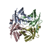
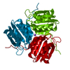
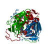
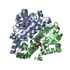
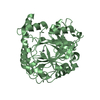
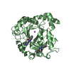
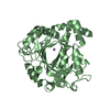
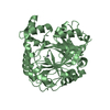
| Title | Crystal structure of raptor adenovirus 1 fibre head, wild-type form | ||||||
 Components Components | FIBER PROTEIN Fibrous protein Fibrous protein | ||||||
 Keywords Keywords | VIRAL PROTEIN | ||||||
| Function / homology |  Function and homology information Function and homology informationadhesion receptor-mediated virion attachment to host cell / viral capsid / cell adhesion / symbiont entry into host cell / host cell nucleusSimilarity search - Function | ||||||
| Biological species |  RAPTOR SIADENOVIRUS A RAPTOR SIADENOVIRUS A | ||||||
| Method | X-RAY DIFFRACTION / SYNCHROTRON / MOLECULAR REPLACEMENT / Resolution: 1.47 Å | ||||||
 Authors Authors | Nguyen, T.H. / van Raaij, M.J. | ||||||
 Citation Citation | Journal: Virol.J. / Year: 2016 Title: Crystal Structure of Raptor Adenovirus 1 Fibre Head and Role of the Beta-Hairpin in Siadenovirus Fibre Head Domains Authors: Nguyen, T.H. / Ballmann, M.Z. / Do, H.T. / Truong, H.N. / Benko, M. / Harrach, B. / van Raaij, M.J. #1: Journal: Infect.Genet.Evol / Year: 2011 Title: Complete Sequence of Raptor Adenovirus 1 Confirms the Characteristic Genome Organization of Siadenoviruses. Authors: Kovacs, E.R. / Benko, M. | ||||||
| History |
|
- Structure visualization
| Structure viewer | Molecule: MolmilJmol/JSmol |
|---|
- Downloads & links
-Download
| PDBx/mmCIF format | 5fjl.cif.gz | 46 KB | Display | PDBx/mmCIF format |
|---|---|---|---|---|
| PDB format | pdb5fjl.ent.gz | 32.4 KB | Display | PDB format |
| PDBx/mmJSON format | 5fjl.json.gz | Tree view | PDBx/mmJSON format | |
| Others |  Other downloads Other downloads |
-Validation report
| Arichive directory | https://data.pdbj.org/pub/pdb/validation_reports/fj/5fjlftp://data.pdbj.org/pub/pdb/validation_reports/fj/5fjl | HTTPS FTP |
|---|
-Related structure data
| Related structure data |  5fldC  3zpeS S: Starting model for refinement C: citing same article ( |
|---|---|
| Similar structure data |
-Links
 PDBj
PDBj
- Assembly
| Deposited unit | 
| ||||||||
|---|---|---|---|---|---|---|---|---|---|
| 1 | 
| ||||||||
| Unit cell |
| ||||||||
| Components on special symmetry positions |
|
-Components
| #1: Protein | Fibrous protein / ADENOVIRUS FIBRE Mass: 19060.756 Da / Num. of mol.: 1 / Fragment: FIBRE HEAD DOMAIN, RESIDUES 324-464 Source method: isolated from a genetically manipulated source Source: (gene. exp.) RAPTOR SIADENOVIRUS A / Description: SEE SECONDARY REFERENCE / Production host:  ESCHERICHIA COLI BL21(DE3) (bacteria) / References: UniProt: F4MI11 ESCHERICHIA COLI BL21(DE3) (bacteria) / References: UniProt: F4MI11 | ||||
|---|---|---|---|---|---|
| #2: Chemical | ChemComp-CL / Chloride  Mass: 35.453 Da / Num. of mol.: 4 / Source method: obtained synthetically / Formula: Cl Mass: 35.453 Da / Num. of mol.: 4 / Source method: obtained synthetically / Formula: Cl#3: Water | ChemComp-HOH / | Water Mass: 18.015 Da / Num. of mol.: 174 / Source method: isolated from a natural source / Formula: H2O Mass: 18.015 Da / Num. of mol.: 174 / Source method: isolated from a natural source / Formula: H2OSequence details | NCBI REFERENCE SEQUENCE YP_004414817.1 | |
-Experimental details
-Experiment
| Experiment | Method: X-RAY DIFFRACTION / Number of used crystals: 1 |
|---|
- Sample preparation
| Crystal | Density Matthews: 2.5 Å3/Da / Density % sol: 51 % / Description: NONE |
|---|---|
| Crystal grow | pH: 9 Details: 20 MM BICINE-NAOH PH 9.0, 50 MM MAGNESIUM CHLORIDE, 5 MM L-ARGININE, 5% (V/V) GLYCEROL, 1.5 M SODIUM CHLORIDE, 10% (V/V) ETHANOL |
-Data collection
| Diffraction | Mean temperature: 100 K |
|---|---|
| Diffraction source | Source: SYNCHROTRON / Site: ALBA  / Beamline: XALOC / Wavelength: 0.97952 / Beamline: XALOC / Wavelength: 0.97952 |
| Detector | Type: DECTRIS PILATUS 6M / Detector: PIXEL / Date: Oct 17, 2013 Details: VERTICAL FOCUSING MIRROR AND HORIZONTAL FOCUSING MIRROR ORTHOGONAL IN A KIRKPATRICK-BAEZ CONFIGURATION |
| Radiation | Monochromator: CRYOGENICALLY COOLED CHANNEL-CUT DCM SI (111) Protocol: SINGLE WAVELENGTH / Monochromatic (M) / Laue (L): M / Scattering type: x-ray |
| Radiation wavelength | Wavelength: 0.97952 Å / Relative weight: 1 |
| Reflection | Resolution: 1.47→47.2 Å / Num. obs: 31365 / % possible obs: 100 % / Redundancy: 9.9 % / Biso Wilson estimate: 17.7 Å2 / Rmerge(I) obs: 0.04 / Net I/σ(I): 29.6 |
| Reflection shell | Resolution: 1.47→1.49 Å / Redundancy: 8.8 % / Rmerge(I) obs: 0.6 / Mean I/σ(I) obs: 3.2 / % possible all: 100 |
- Processing
| Software |
| ||||||||||||||||||||||||||||||||||||||||||||||||||||||||||||||||||||||||||||||||||||||||||||||||||||||||||||||||||||||||||||||||||||||||||||||||||||||||||||||||||||||||||||||||||||||
|---|---|---|---|---|---|---|---|---|---|---|---|---|---|---|---|---|---|---|---|---|---|---|---|---|---|---|---|---|---|---|---|---|---|---|---|---|---|---|---|---|---|---|---|---|---|---|---|---|---|---|---|---|---|---|---|---|---|---|---|---|---|---|---|---|---|---|---|---|---|---|---|---|---|---|---|---|---|---|---|---|---|---|---|---|---|---|---|---|---|---|---|---|---|---|---|---|---|---|---|---|---|---|---|---|---|---|---|---|---|---|---|---|---|---|---|---|---|---|---|---|---|---|---|---|---|---|---|---|---|---|---|---|---|---|---|---|---|---|---|---|---|---|---|---|---|---|---|---|---|---|---|---|---|---|---|---|---|---|---|---|---|---|---|---|---|---|---|---|---|---|---|---|---|---|---|---|---|---|---|---|---|---|---|
| Refinement | Method to determine structure: MOLECULAR REPLACEMENT Starting model: PDB ENTRY 3ZPE Resolution: 1.47→45 Å / Cor.coef. Fo:Fc: 0.975 / Cor.coef. Fo:Fc free: 0.971 / SU B: 0.93 / SU ML: 0.036 / Cross valid method: THROUGHOUT / ESU R: 0.054 / ESU R Free: 0.056 / Stereochemistry target values: MAXIMUM LIKELIHOOD Details: HYDROGENS HAVE BEEN ADDED IN THE RIDING POSITIONS. U VALUES REFINED INDIVIDUALLY
| ||||||||||||||||||||||||||||||||||||||||||||||||||||||||||||||||||||||||||||||||||||||||||||||||||||||||||||||||||||||||||||||||||||||||||||||||||||||||||||||||||||||||||||||||||||||
| Solvent computation | Ion probe radii: 0.8 Å / Shrinkage radii: 0.8 Å / VDW probe radii: 1.2 Å / Solvent model: MASK | ||||||||||||||||||||||||||||||||||||||||||||||||||||||||||||||||||||||||||||||||||||||||||||||||||||||||||||||||||||||||||||||||||||||||||||||||||||||||||||||||||||||||||||||||||||||
| Displacement parameters | Biso mean: 24.629 Å2
| ||||||||||||||||||||||||||||||||||||||||||||||||||||||||||||||||||||||||||||||||||||||||||||||||||||||||||||||||||||||||||||||||||||||||||||||||||||||||||||||||||||||||||||||||||||||
| Refinement step | Cycle: LAST / Resolution: 1.47→45 Å
| ||||||||||||||||||||||||||||||||||||||||||||||||||||||||||||||||||||||||||||||||||||||||||||||||||||||||||||||||||||||||||||||||||||||||||||||||||||||||||||||||||||||||||||||||||||||
| Refine LS restraints |
|