 Movie
Movie Controller

[English] 日本語
 Yorodumi
- PDB-5fhw: Hen Egg-White Lysozyme (HEWL) complexed with Hf(IV)-substituted W... -
+ Open data

- Basic information
| Entry | Database: PDB / ID: 5fhw | ||||||
|---|---|---|---|---|---|---|---|
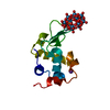
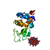
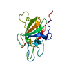
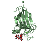
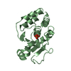
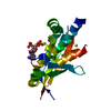
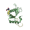
| Title | Hen Egg-White Lysozyme (HEWL) complexed with Hf(IV)-substituted Wells Dawson-type polyoxometalate | ||||||
 Components Components | Lysozyme C | ||||||
 Keywords Keywords |  HYDROLASE / Complex / polyoxometalate / hafnium / Wells Dawson HYDROLASE / Complex / polyoxometalate / hafnium / Wells Dawson | ||||||
| Function / homology |  Function and homology information Function and homology informationLactose synthesis / Antimicrobial peptides / Neutrophil degranulation / beta-N-acetylglucosaminidase activity / cell wall macromolecule catabolic process / lysozyme / lysozyme activity / killing of cells of another organism / defense response to Gram-negative bacterium / defense response to Gram-positive bacterium ...Lactose synthesis / Antimicrobial peptides / Neutrophil degranulation / beta-N-acetylglucosaminidase activity / cell wall macromolecule catabolic process / lysozyme / lysozyme activity / killing of cells of another organism / defense response to Gram-negative bacterium / defense response to Gram-positive bacterium / defense response to bacterium / endoplasmic reticulum / extracellular space / identical protein binding / cytoplasmSimilarity search - Function | ||||||
| Biological species |  Gallus gallus (chicken) Gallus gallus (chicken) | ||||||
| Method | X-RAY DIFFRACTION / SYNCHROTRON / MOLECULAR REPLACEMENT / molecular replacement / Resolution: 1.52 Å | ||||||
 Authors Authors | Vandebroek, L. / De Zitter, E. / Van Meervelt, L. | ||||||
 Citation Citation | Journal: To Be Published Title: Structural Characterization of the Complex between Hen Egg- White Lysozyme and HfIV-Substituted Wells Dawson Polyoxometalate as Artificial Protease Authors: Vandebroek, L. / De Zitter, E. / Van Meervelt, L. / Parac-Vogt, T.N. | ||||||
| History |
|
- Structure visualization
| Structure viewer | Molecule: MolmilJmol/JSmol |
|---|
- Downloads & links
-Download
| PDBx/mmCIF format | 5fhw.cif.gz | 47.4 KB | Display | PDBx/mmCIF format |
|---|---|---|---|---|
| PDB format | pdb5fhw.ent.gz | 31.5 KB | Display | PDB format |
| PDBx/mmJSON format | 5fhw.json.gz | Tree view | PDBx/mmJSON format | |
| Others |  Other downloads Other downloads |
-Validation report
| Arichive directory | https://data.pdbj.org/pub/pdb/validation_reports/fh/5fhwftp://data.pdbj.org/pub/pdb/validation_reports/fh/5fhw | HTTPS FTP |
|---|
-Related structure data
| Related structure data | 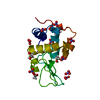 2vb1S S: Starting model for refinement |
|---|---|
| Similar structure data |


-Links
 PDBj
PDBj


- Assembly
| Deposited unit | 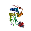
| |||||||||
|---|---|---|---|---|---|---|---|---|---|---|
| 1 |
| |||||||||
| Unit cell |
| |||||||||
| Components on special symmetry positions |
|
-Components
| #1: Protein | Mass: 14331.160 Da / Num. of mol.: 1 / Source method: isolated from a natural source / Source: (natural) Gallus gallus (chicken) / References: UniProt: P00698, lysozyme | ||
|---|---|---|---|
| #2: Chemical | ChemComp-HFW /  Mass: 4343.697 Da / Num. of mol.: 1 / Source method: obtained synthetically / Formula: HfO61P2W17 Mass: 4343.697 Da / Num. of mol.: 1 / Source method: obtained synthetically / Formula: HfO61P2W17 | ||
| #3: Chemical |   Mass: 39.098 Da / Num. of mol.: 2 / Source method: obtained synthetically / Formula: K Mass: 39.098 Da / Num. of mol.: 2 / Source method: obtained synthetically / Formula: K#4: Water | ChemComp-HOH / | Water Mass: 18.015 Da / Num. of mol.: 172 / Source method: isolated from a natural source / Formula: H2O Mass: 18.015 Da / Num. of mol.: 172 / Source method: isolated from a natural source / Formula: H2O |
-Experimental details
-Experiment
| Experiment | Method: X-RAY DIFFRACTION / Number of used crystals: 1 |
|---|
- Sample preparation
| Crystal | Density Matthews: 2 Å3/Da / Density % sol: 38.45 % |
|---|---|
| Crystal grow | Temperature: 289 K / Method: vapor diffusion, sitting drop / pH: 6.5 Details: 25 mg/mL Lysozyme C, 1.45 mM 1:2 Hf-Wells Dawson, 2.0 M sodium chloride, 0.1 M potassium phosphate monobasic, 0.1 M sodium phosphate monobasic monohydrate, 0.1 M MES pH = 6.5 |
-Data collection
| Diffraction | Mean temperature: 100 K | |||||||||||||||||||||||||||
|---|---|---|---|---|---|---|---|---|---|---|---|---|---|---|---|---|---|---|---|---|---|---|---|---|---|---|---|---|
| Diffraction source | Source: SYNCHROTRON / Site: SLS  / Beamline: X06SA / Wavelength: 1 Å / Beamline: X06SA / Wavelength: 1 Å | |||||||||||||||||||||||||||
| Detector | Type: DECTRIS PILATUS 2M / Detector: PIXEL / Date: May 23, 2015 | |||||||||||||||||||||||||||
| Radiation | Protocol: SINGLE WAVELENGTH / Monochromatic (M) / Laue (L): M / Scattering type: x-ray | |||||||||||||||||||||||||||
| Radiation wavelength | Wavelength: 1 Å / Relative weight: 1 | |||||||||||||||||||||||||||
| Reflection | Resolution: 1.52→39.48 Å / Num. all: 130169 / Num. obs: 18463 / % possible obs: 100 % / Redundancy: 7.1 % / Biso Wilson estimate: 10.3 Å2 / CC1/2: 0.998 / Rmerge(I) obs: 0.12 / Rpim(I) all: 0.048 / Net I/σ(I): 17.3 / Num. measured all: 130169 | |||||||||||||||||||||||||||
| Reflection shell | Diffraction-ID: 1 / Rejects: 0
|
-Phasing
| Phasing | Method: molecular replacement | |||||||||
|---|---|---|---|---|---|---|---|---|---|---|
| Phasing MR |
|
- Processing
| Software |
| |||||||||||||||||||||||||||||||||||||||||||||||||
|---|---|---|---|---|---|---|---|---|---|---|---|---|---|---|---|---|---|---|---|---|---|---|---|---|---|---|---|---|---|---|---|---|---|---|---|---|---|---|---|---|---|---|---|---|---|---|---|---|---|---|
| Refinement | Method to determine structure: MOLECULAR REPLACEMENT Starting model: 2VB1 Resolution: 1.52→35.308 Å / SU ML: 0.11 / Cross valid method: FREE R-VALUE / σ(F): 1.34 / Phase error: 20.62 / Stereochemistry target values: ML
| |||||||||||||||||||||||||||||||||||||||||||||||||
| Solvent computation | Shrinkage radii: 0.9 Å / VDW probe radii: 1.11 Å / Solvent model: FLAT BULK SOLVENT MODEL | |||||||||||||||||||||||||||||||||||||||||||||||||
| Displacement parameters | Biso max: 47.97 Å2 / Biso mean: 19.2414 Å2 / Biso min: 9.1 Å2 | |||||||||||||||||||||||||||||||||||||||||||||||||
| Refinement step | Cycle: final / Resolution: 1.52→35.308 Å
| |||||||||||||||||||||||||||||||||||||||||||||||||
| Refine LS restraints |
| |||||||||||||||||||||||||||||||||||||||||||||||||
| LS refinement shell | Refine-ID: X-RAY DIFFRACTION / Total num. of bins used: 6
|
