 Movie
Movie Controller

[English] 日本語
 Yorodumi
- PDB-5fbz: Structure of subtilase SubHal from Bacillus halmapalus - complex ... -
+ Open data

- Basic information
| Entry | Database: PDB / ID: 5fbz | ||||||
|---|---|---|---|---|---|---|---|
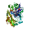
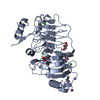
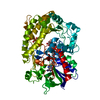
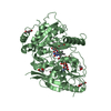
| Title | Structure of subtilase SubHal from Bacillus halmapalus - complex with chymotrypsin inhibitor CI2A | ||||||
 Components Components |
| ||||||
 Keywords Keywords |  HYDROLASE / Protease / Subtilase / Calcium binding / CI2A inhibitor HYDROLASE / Protease / Subtilase / Calcium binding / CI2A inhibitor | ||||||
| Function / homology |  Function and homology information Function and homology information3.4.21.14 / serine-type endopeptidase inhibitor activity / response to wounding / serine-type endopeptidase activity / proteolysis / metal ion bindingSimilarity search - Function | ||||||
| Biological species |  Bacillus halmapalus (bacteria) Bacillus halmapalus (bacteria) Hordeum vulgare (barley) Hordeum vulgare (barley) | ||||||
| Method | X-RAY DIFFRACTION / MIR / Resolution: 1.9 Å | ||||||
 Authors Authors | Dohnalek, J. / Brzozowski, A.M. / Svendsen, A. / Wilson, K.S. | ||||||
 Citation Citation | Book title: Understanding enzymes; Function, Design, Engineering and Analysis Journal: Book / Year: 2016 Title: Stabilization of Enzymes by Metal Binding: Structures of Two Alkalophilic Bacillus Subtilases and Analysis of the Second Metal-Binding Site of the Subtilase Family Authors: Dohnalek, J. / McAuley, K.E. / Brzozowski, A.M. / Oestergaard, P.R. / Svendsen, A. / Wilson, K.S. | ||||||
| History |
|
- Structure visualization
| Structure viewer | Molecule: MolmilJmol/JSmol |
|---|
- Downloads & links
-Download
| PDBx/mmCIF format | 5fbz.cif.gz | 221.8 KB | Display | PDBx/mmCIF format |
|---|---|---|---|---|
| PDB format | pdb5fbz.ent.gz | 180.9 KB | Display | PDB format |
| PDBx/mmJSON format | 5fbz.json.gz | Tree view | PDBx/mmJSON format | |
| Others |  Other downloads Other downloads |
-Validation report
| Arichive directory | https://data.pdbj.org/pub/pdb/validation_reports/fb/5fbzftp://data.pdbj.org/pub/pdb/validation_reports/fb/5fbz | HTTPS FTP |
|---|
-Related structure data


-Links
 PDBj
PDBj

- Assembly
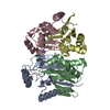
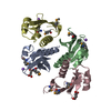
| Deposited unit | 
| ||||||||
|---|---|---|---|---|---|---|---|---|---|
| 1 | 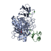
| ||||||||
| 2 | 
| ||||||||
| 3 | 
| ||||||||
| Unit cell |
|
-Components
| #1: Protein | Mass: 45340.984 Da / Num. of mol.: 2 Source method: isolated from a genetically manipulated source Details: The N-terminal asparagine is carbamoylated to N-carboxyasparagine, The sequence is available in patent WO 2004083362 Source: (gene. exp.) Bacillus halmapalus (bacteria) / Production host: Bacillus subtilis (bacteria) / References: UniProt: A0A182DWC7*PLUS, 3.4.21.14#2: Protein | Mass: 8107.385 Da / Num. of mol.: 2 / Fragment: UNP residues 13-84 Source method: isolated from a genetically manipulated source Source: (gene. exp.) Hordeum vulgare (barley) / Production host: Escherichia coli (E. coli) / References: UniProt: P01053#3: Protein/peptide | | Mass: 557.703 Da / Num. of mol.: 1 Source method: isolated from a genetically manipulated source Details: The fragment was produced probably during crystallization. The fragment length is not known. Source: (gene. exp.) Bacillus halmapalus (bacteria) / Production host: Bacillus subtilis (bacteria) / References: 3.4.21.14#4: Chemical | ChemComp-CA /   Mass: 40.078 Da / Num. of mol.: 6 / Source method: obtained synthetically / Formula: Ca Mass: 40.078 Da / Num. of mol.: 6 / Source method: obtained synthetically / Formula: Ca#5: Water | ChemComp-HOH / | Water Mass: 18.015 Da / Num. of mol.: 1098 / Source method: isolated from a natural source / Formula: H2O Mass: 18.015 Da / Num. of mol.: 1098 / Source method: isolated from a natural source / Formula: H2O |
|---|
-Experimental details
-Experiment
| Experiment | Method: X-RAY DIFFRACTION |
|---|
- Sample preparation
| Crystal | Density Matthews: 2.38 Å3/Da / Density % sol: 48.4 % / Description: Plate-like crystal |
|---|---|
| Crystal grow | Temperature: 291 K / Method: vapor diffusion, hanging drop / pH: 7.5 Details: Plate-like crystals were grown by hanging drop vapour diffusion with the drop consisting of 2 microliters of 15-20 mg/mL concentration protein, 10 mM sodium cacodylate in HCl buffer, pH 6.5 ...Details: Plate-like crystals were grown by hanging drop vapour diffusion with the drop consisting of 2 microliters of 15-20 mg/mL concentration protein, 10 mM sodium cacodylate in HCl buffer, pH 6.5 and 1 microliter of reservoir solution: 20% w/v PEG 4000, 0.1 M HEPES buffer, pH 7.5, 10% v/v isopropanol. |
-Data collection
| Diffraction | Mean temperature: 120 K |
|---|---|
| Diffraction source | Source: ROTATING ANODE / Type: RIGAKU RU300 / Wavelength: 1.54178 Å |
| Detector | Type: MAR scanner 345 mm plate / Detector: IMAGE PLATE / Date: Jun 9, 1998 |
| Radiation | Protocol: SINGLE WAVELENGTH / Monochromatic (M) / Laue (L): M / Scattering type: x-ray |
| Radiation wavelength | Wavelength: 1.54178 Å / Relative weight: 1 |
| Reflection | Resolution: 1.9→20 Å / Num. all: 59477 / Num. obs: 59477 / % possible obs: 76.6 % / Observed criterion σ(I): -3 / Redundancy: 9.2 % / Biso Wilson estimate: 25.5 Å2 / Rmerge(I) obs: 0.046 / Net I/σ(I): 22 |
| Reflection shell | Resolution: 1.9→1.93 Å / Rmerge(I) obs: 0.128 / Mean I/σ(I) obs: 5.4 / % possible all: 14.5 |
- Processing
| Software |
| ||||||||||||||||||||||||||||||||||||||||||||||||||||||||||||||||||||||||||||||||||||||||||||||||||||||||||||||||||||||||||||||||||||||||||||||||||||||||||||||||||||||||||||||||||||||
|---|---|---|---|---|---|---|---|---|---|---|---|---|---|---|---|---|---|---|---|---|---|---|---|---|---|---|---|---|---|---|---|---|---|---|---|---|---|---|---|---|---|---|---|---|---|---|---|---|---|---|---|---|---|---|---|---|---|---|---|---|---|---|---|---|---|---|---|---|---|---|---|---|---|---|---|---|---|---|---|---|---|---|---|---|---|---|---|---|---|---|---|---|---|---|---|---|---|---|---|---|---|---|---|---|---|---|---|---|---|---|---|---|---|---|---|---|---|---|---|---|---|---|---|---|---|---|---|---|---|---|---|---|---|---|---|---|---|---|---|---|---|---|---|---|---|---|---|---|---|---|---|---|---|---|---|---|---|---|---|---|---|---|---|---|---|---|---|---|---|---|---|---|---|---|---|---|---|---|---|---|---|---|---|
| Refinement | Method to determine structure: MIR / Resolution: 1.9→19.97 Å / Cor.coef. Fo:Fc: 0.966 / SU B: 1.525 / SU ML: 0.046 / Cross valid method: FREE R-VALUE / ESU R: 0.159 / Stereochemistry target values: MAXIMUM LIKELIHOOD / Details: HYDROGENS HAVE BEEN ADDED IN THE RIDING POSITIONS
| ||||||||||||||||||||||||||||||||||||||||||||||||||||||||||||||||||||||||||||||||||||||||||||||||||||||||||||||||||||||||||||||||||||||||||||||||||||||||||||||||||||||||||||||||||||||
| Solvent computation | Ion probe radii: 0.8 Å / Shrinkage radii: 0.8 Å / VDW probe radii: 1.2 Å / Solvent model: MASK | ||||||||||||||||||||||||||||||||||||||||||||||||||||||||||||||||||||||||||||||||||||||||||||||||||||||||||||||||||||||||||||||||||||||||||||||||||||||||||||||||||||||||||||||||||||||
| Displacement parameters | Biso mean: 14.619 Å2
| ||||||||||||||||||||||||||||||||||||||||||||||||||||||||||||||||||||||||||||||||||||||||||||||||||||||||||||||||||||||||||||||||||||||||||||||||||||||||||||||||||||||||||||||||||||||
| Refinement step | Cycle: 1 / Resolution: 1.9→19.97 Å
| ||||||||||||||||||||||||||||||||||||||||||||||||||||||||||||||||||||||||||||||||||||||||||||||||||||||||||||||||||||||||||||||||||||||||||||||||||||||||||||||||||||||||||||||||||||||
| Refine LS restraints |
|