 Movie
Movie Controller


+ Open data

- Basic information
| Entry | Database: PDB / ID: 5fb8 | ||||||
|---|---|---|---|---|---|---|---|
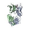
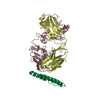
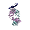
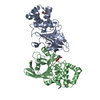
| Title | Structure of Interleukin-16 bound to the 14.1 antibody | ||||||
 Components Components |
| ||||||
 Keywords Keywords |  IMMUNE SYSTEM / Cytokine / Interleukin / Antibody / Complex IMMUNE SYSTEM / Cytokine / Interleukin / Antibody / Complex | ||||||
| Function / homology |  Function and homology information Function and homology informationpositive regulation of interleukin-1 alpha production / induction of positive chemotaxis / CD4 receptor binding / Flemming body / leukocyte chemotaxis / Other interleukin signaling / regulation of calcium ion transport / positive regulation of interleukin-12 production / cytokine activity / positive regulation of inflammatory response ...positive regulation of interleukin-1 alpha production / induction of positive chemotaxis / CD4 receptor binding / Flemming body / leukocyte chemotaxis / Other interleukin signaling / regulation of calcium ion transport / positive regulation of interleukin-12 production / cytokine activity / positive regulation of inflammatory response / positive regulation of interleukin-6 production / nuclear speck / immune response / focal adhesion / extracellular space / extracellular region / plasma membrane / cytosolSimilarity search - Function | ||||||
| Biological species |  Mus musculus (house mouse) Mus musculus (house mouse) Homo sapiens (human) Homo sapiens (human) | ||||||
| Method | X-RAY DIFFRACTION / SYNCHROTRON / MOLECULAR REPLACEMENT / Resolution: 2.07 Å | ||||||
 Authors Authors | Hall, G. / Cowan, R. / Bayliss, R. / Carr, M. | ||||||
 Citation Citation | Journal: J.Biol.Chem. / Year: 2016 Title: Structure of a Potential Therapeutic Antibody Bound to Interleukin-16 (IL-16): MECHANISTIC INSIGHTS AND NEW THERAPEUTIC OPPORTUNITIES. Authors: Hall, G. / Cullen, E. / Sawmynaden, K. / Arnold, J. / Fox, S. / Cowan, R. / Muskett, F.W. / Matthews, D. / Merritt, A. / Kettleborough, C. / Cruikshank, W. / Taylor, D. / Bayliss, R. / Carr, M.D. | ||||||
| History |
|
- Structure visualization
| Structure viewer | Molecule: MolmilJmol/JSmol |
|---|
- Downloads & links
-Download
| PDBx/mmCIF format | 5fb8.cif.gz | 124.5 KB | Display | PDBx/mmCIF format |
|---|---|---|---|---|
| PDB format | pdb5fb8.ent.gz | 92.9 KB | Display | PDB format |
| PDBx/mmJSON format | 5fb8.json.gz | Tree view | PDBx/mmJSON format | |
| Others |  Other downloads Other downloads |
-Validation report
| Arichive directory | https://data.pdbj.org/pub/pdb/validation_reports/fb/5fb8ftp://data.pdbj.org/pub/pdb/validation_reports/fb/5fb8 | HTTPS FTP |
|---|
-Related structure data


-Links
 PDBj
PDBj


- Assembly
| Deposited unit | 
| ||||||||
|---|---|---|---|---|---|---|---|---|---|
| 1 |
| ||||||||
| Unit cell |
|
-Components
-Protein , 1 types, 1 molecules C
| #3: Protein | Mass: 10785.303 Da / Num. of mol.: 1 / Fragment: UNP residues 1224-1323 Source method: isolated from a genetically manipulated source Source: (gene. exp.) Homo sapiens (human) / Gene: IL16 / Plasmid: pLEICS-01 / Production host:  Escherichia coli BL21(DE3) (bacteria) / References: UniProt: Q14005 Escherichia coli BL21(DE3) (bacteria) / References: UniProt: Q14005 |
|---|
-Antibody , 2 types, 2 molecules AB
| #1: Antibody | Mass: 23818.469 Da / Num. of mol.: 1 Source method: isolated from a genetically manipulated source Source: (gene. exp.) Mus musculus (house mouse) / Plasmid: pCMV / Cell line (production host): Expi293F / Production host: Homo sapiens (human) |
|---|---|
| #2: Antibody | Mass: 24181.986 Da / Num. of mol.: 1 Source method: isolated from a genetically manipulated source Source: (gene. exp.) Mus musculus (house mouse) / Plasmid: pCMV / Cell line (production host): Expi293F / Production host: Homo sapiens (human) |
-Non-polymers , 4 types, 302 molecules 


| #4: Chemical | Sulfate Mass: 96.063 Da / Num. of mol.: 3 / Source method: obtained synthetically / Formula: SO4 Mass: 96.063 Da / Num. of mol.: 3 / Source method: obtained synthetically / Formula: SO4#5: Chemical | ChemComp-EDO / Ethylene glycol Mass: 62.068 Da / Num. of mol.: 7 / Source method: obtained synthetically / Formula: C2H6O2 Mass: 62.068 Da / Num. of mol.: 7 / Source method: obtained synthetically / Formula: C2H6O2#6: Chemical | ChemComp-TRS / | Tris Mass: 122.143 Da / Num. of mol.: 1 / Source method: obtained synthetically / Formula: C4H12NO3 / Comment: pH buffer*YM Mass: 122.143 Da / Num. of mol.: 1 / Source method: obtained synthetically / Formula: C4H12NO3 / Comment: pH buffer*YM#7: Water | ChemComp-HOH / | WaterMass: 18.015 Da / Num. of mol.: 291 / Source method: isolated from a natural source / Formula: H2O |
|---|
-Experimental details
-Experiment
| Experiment | Method: X-RAY DIFFRACTION |
|---|
- Sample preparation
| Crystal | Density Matthews: 3.29 Å3/Da / Density % sol: 62.6 % |
|---|---|
| Crystal grow | Temperature: 293 K / Method: vapor diffusion, hanging drop / pH: 6.5 Details: 16% PEG3350, 0.1 M Bis-Tris-Propane, pH 6.5 and 0.2 M sodium sulphate. |
-Data collection
| Diffraction | Mean temperature: 100 K |
|---|---|
| Diffraction source | Source: SYNCHROTRON / Site: Diamond  / Beamline: I03 / Wavelength: 0.97626 Å / Beamline: I03 / Wavelength: 0.97626 Å |
| Detector | Type: DECTRIS PILATUS3 6M / Detector: PIXEL / Date: May 3, 2013 |
| Radiation | Protocol: SINGLE WAVELENGTH / Monochromatic (M) / Laue (L): M / Scattering type: x-ray |
| Radiation wavelength | Wavelength: 0.97626 Å / Relative weight: 1 |
| Reflection | Resolution: 2.07→29.45 Å / Num. obs: 48100 / % possible obs: 99.81 % / Redundancy: 6.3 % / Rmerge(I) obs: 0.055 / Net I/σ(I): 17.79 |
| Reflection shell | Resolution: 2.07→2.144 Å / Redundancy: 6.5 % / Rmerge(I) obs: 0.695 / Mean I/σ(I) obs: 3.44 / % possible all: 99.85 |
- Processing
| Software |
| ||||||||||||||||||||||||||||||||||||||||||||||||||||||||||||||||||||||||||||||||||||||||||||||||||||||||||||||||||||||||||||||
|---|---|---|---|---|---|---|---|---|---|---|---|---|---|---|---|---|---|---|---|---|---|---|---|---|---|---|---|---|---|---|---|---|---|---|---|---|---|---|---|---|---|---|---|---|---|---|---|---|---|---|---|---|---|---|---|---|---|---|---|---|---|---|---|---|---|---|---|---|---|---|---|---|---|---|---|---|---|---|---|---|---|---|---|---|---|---|---|---|---|---|---|---|---|---|---|---|---|---|---|---|---|---|---|---|---|---|---|---|---|---|---|---|---|---|---|---|---|---|---|---|---|---|---|---|---|---|---|
| Refinement | Method to determine structure: MOLECULAR REPLACEMENT Starting model: 1I7Z for chain A, 4F33 for chain B, and 1I16 for chain C Resolution: 2.07→29.45 Å / SU ML: 0.22 / Cross valid method: FREE R-VALUE / σ(F): 1.34 / Phase error: 21.67 / Stereochemistry target values: ML
| ||||||||||||||||||||||||||||||||||||||||||||||||||||||||||||||||||||||||||||||||||||||||||||||||||||||||||||||||||||||||||||||
| Solvent computation | Shrinkage radii: 0.9 Å / VDW probe radii: 1.11 Å / Solvent model: FLAT BULK SOLVENT MODEL | ||||||||||||||||||||||||||||||||||||||||||||||||||||||||||||||||||||||||||||||||||||||||||||||||||||||||||||||||||||||||||||||
| Refinement step | Cycle: LAST / Resolution: 2.07→29.45 Å
| ||||||||||||||||||||||||||||||||||||||||||||||||||||||||||||||||||||||||||||||||||||||||||||||||||||||||||||||||||||||||||||||
| Refine LS restraints |
| ||||||||||||||||||||||||||||||||||||||||||||||||||||||||||||||||||||||||||||||||||||||||||||||||||||||||||||||||||||||||||||||
| LS refinement shell |
|
