- PDB-3s3h: Crystal structure of the catalytic domain of PTP10D from Drosophi... -
+
Open data
ID or keywords:
Loading...
-
Basic information
Entry
Database: PDB / ID: 3s3h
Title
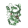
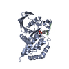
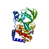
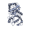
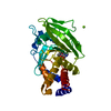
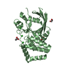
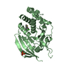
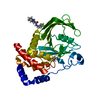
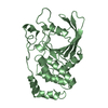
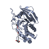
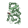
Crystal structure of the catalytic domain of PTP10D from Drosophila melanogaster with a phosphopeptide substrate GP4
Components
Tyrosine-protein phosphatase 10D
phosphopeptide GP4
Keywords
HYDROLASE / Differentiation / Neurogenesis / Signal transduction / developmental Protein / Protein Phosphatase / Protein Tyrosine Phosphatase
Function / homology
Function and homology information
branching involved in open tracheal system development / open tracheal system development / negative regulation of fibroblast growth factor receptor signaling pathway / Neutrophil degranulation / transmembrane receptor protein tyrosine phosphatase activity / motor neuron axon guidance / negative regulation of epidermal growth factor receptor signaling pathway / negative regulation of vascular endothelial growth factor receptor signaling pathway / long-term memory / protein dephosphorylation ...branching involved in open tracheal system development / open tracheal system development / negative regulation of fibroblast growth factor receptor signaling pathway / Neutrophil degranulation / transmembrane receptor protein tyrosine phosphatase activity / motor neuron axon guidance / negative regulation of epidermal growth factor receptor signaling pathway / negative regulation of vascular endothelial growth factor receptor signaling pathway / long-term memory / protein dephosphorylation / protein-tyrosine-phosphatase / central nervous system development / protein tyrosine phosphatase activity / axon guidance / apical part of cell / axon / membrane Similarity search - Function
PTPRJ, transmembrane domain / TM proximal of protein tyrosine phosphatase, receptor type J / Protein tyrosine phosphatase superfamily / Protein-Tyrosine Phosphatase; Chain A / Protein tyrosine phosphatase, catalytic domain / PTP type protein phosphatase domain profile. / Protein-tyrosine phosphatase / Tyrosine-specific protein phosphatase, PTPase domain / Protein-tyrosine phosphatase, catalytic / Protein tyrosine phosphatase, catalytic domain motif ...PTPRJ, transmembrane domain / TM proximal of protein tyrosine phosphatase, receptor type J / Protein tyrosine phosphatase superfamily / Protein-Tyrosine Phosphatase; Chain A / Protein tyrosine phosphatase, catalytic domain / PTP type protein phosphatase domain profile. / Protein-tyrosine phosphatase / Tyrosine-specific protein phosphatase, PTPase domain / Protein-tyrosine phosphatase, catalytic / Protein tyrosine phosphatase, catalytic domain motif / Tyrosine specific protein phosphatases active site. / Protein-tyrosine phosphatase, active site / Tyrosine-specific protein phosphatases domain / Tyrosine specific protein phosphatases domain profile. / Protein-tyrosine phosphatase-like / Fibronectin type III domain / Fibronectin type 3 domain / Fibronectin type-III domain profile. / Fibronectin type III / Fibronectin type III superfamily / Immunoglobulin-like fold / Alpha-Beta Complex / Alpha Beta Similarity search - Domain/homology
In the structure databanks used in Yorodumi, some data are registered as the other names, "COVID-19 virus" and "2019-nCoV". Here are the details of the virus and the list of structure data.
Jan 31, 2019. EMDB accession codes are about to change! (news from PDBe EMDB page)
EMDB accession codes are about to change! (news from PDBe EMDB page)
The allocation of 4 digits for EMDB accession codes will soon come to an end. Whilst these codes will remain in use, new EMDB accession codes will include an additional digit and will expand incrementally as the available range of codes is exhausted. The current 4-digit format prefixed with “EMD-” (i.e. EMD-XXXX) will advance to a 5-digit format (i.e. EMD-XXXXX), and so on. It is currently estimated that the 4-digit codes will be depleted around Spring 2019, at which point the 5-digit format will come into force.
The EM Navigator/Yorodumi systems omit the EMD- prefix.
Related info.:Q: What is EMD? / ID/Accession-code notation in Yorodumi/EM Navigator
Yorodumi is a browser for structure data from EMDB, PDB, SASBDB, etc.
This page is also the successor to EM Navigator detail page, and also detail information page/front-end page for Omokage search.
The word "yorodu" (or yorozu) is an old Japanese word meaning "ten thousand". "mi" (miru) is to see.
Related info.:EMDB / PDB / SASBDB / Comparison of 3 databanks / Yorodumi Search / Aug 31, 2016. New EM Navigator & Yorodumi / Yorodumi Papers / Jmol/JSmol / Function and homology information / Changes in new EM Navigator and Yorodumi
 Movie
Movie Controller

 Yorodumi
 Open data
 Basic information
 Components
 Keywords
 HYDROLASE / Differentiation /
  Function and homology information
 Authors
 Citation
 Structure visualization
 Downloads & links
 Other downloads
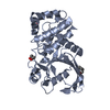


 PDBj
PDBj


 Assembly
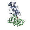

 Mass: 74.122 Da / Num. of mol.: 1 / Source method: obtained synthetically / Formula: C4H10O
 Mass: 90.121 Da / Num. of mol.: 1 / Source method: obtained synthetically / Formula: C4H10O2
 Mass: 18.015 Da / Num. of mol.: 82 / Source method: isolated from a natural source / Formula: H2O
 Sample preparation
 / Beamline: BM14 / Wavelength: 1
  Processing