 Movie
Movie Controller


+ Open data

- Basic information
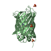
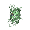
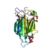
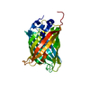
| Entry | Database: PDB / ID: 5f9p | ||||||
|---|---|---|---|---|---|---|---|
| Title | Crystal structure study of anthrone oxidase-like protein | ||||||
 Components Components | Anthrone oxidase-like protein | ||||||
 Keywords Keywords |  OXIDOREDUCTASE / anthrone oxidase-like protein / oxidative ring-B opening / activity site OXIDOREDUCTASE / anthrone oxidase-like protein / oxidative ring-B opening / activity site | ||||||
| Function / homology | Antibiotic biosynthesis monooxygenase / Antibiotic biosynthesis monooxygenase domain / Dimeric alpha-beta barrel / Anthrone oxidase-like protein Function and homology information Function and homology information | ||||||
| Biological species |  Streptomyces ambofaciens (bacteria) Streptomyces ambofaciens (bacteria) | ||||||
| Method | X-RAY DIFFRACTION / SYNCHROTRON / SAD / Resolution: 2.078 Å | ||||||
 Authors Authors | Gao, X. / Wu, D. / Fan, K. / Liu, Z.-J. | ||||||
 Citation Citation | Journal: ACS Chem. Biol. / Year: 2017 Title: Structure and Function of a C-C Bond Cleaving Oxygenase in Atypical Angucycline Biosynthesis Authors: Pan, G. / Gao, X. / Fan, K. / Liu, J. / Meng, B. / Gao, J. / Wang, B. / Zhang, C. / Han, H. / Ai, G. / Chen, Y. / Wu, D. / Liu, Z.J. / Yang, K. | ||||||
| History |
|
- Structure visualization
| Structure viewer | Molecule: MolmilJmol/JSmol |
|---|
- Downloads & links
-Download
| PDBx/mmCIF format | 5f9p.cif.gz | 106.7 KB | Display | PDBx/mmCIF format |
|---|---|---|---|---|
| PDB format | pdb5f9p.ent.gz | 87.2 KB | Display | PDB format |
| PDBx/mmJSON format | 5f9p.json.gz | Tree view | PDBx/mmJSON format | |
| Others |  Other downloads Other downloads |
-Validation report
| Arichive directory | https://data.pdbj.org/pub/pdb/validation_reports/f9/5f9pftp://data.pdbj.org/pub/pdb/validation_reports/f9/5f9p | HTTPS FTP |
|---|
-Related structure data
| Similar structure data |
|---|


-Links
 PDBj
PDBj- Assembly
| Deposited unit | 
| ||||||||
|---|---|---|---|---|---|---|---|---|---|
| 1 | 
| ||||||||
| 2 | 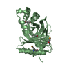
| ||||||||
| Unit cell |
| ||||||||
| Components on special symmetry positions |
|
-Components
| #1: Protein | Mass: 26746.357 Da / Num. of mol.: 2 Source method: isolated from a genetically manipulated source Source: (gene. exp.) Streptomyces ambofaciens (bacteria) / Gene: alpJ, DSMT0187 / Plasmid: pET-30a / Production host: Escherichia coli BL21(DE3) (bacteria) / Strain (production host): BL21 (DE3) / References: UniProt: Q6VMI5#2: Chemical | Glycerol  Mass: 92.094 Da / Num. of mol.: 2 / Source method: obtained synthetically / Formula: C3H8O3 Mass: 92.094 Da / Num. of mol.: 2 / Source method: obtained synthetically / Formula: C3H8O3#3: Water | ChemComp-HOH / | Water Mass: 18.015 Da / Num. of mol.: 312 / Source method: isolated from a natural source / Formula: H2O Mass: 18.015 Da / Num. of mol.: 312 / Source method: isolated from a natural source / Formula: H2O |
|---|
-Experimental details
-Experiment
| Experiment | Method: X-RAY DIFFRACTION / Number of used crystals: 1 |
|---|
- Sample preparation
| Crystal | Density Matthews: 2.1 Å3/Da / Density % sol: 41.56 % |
|---|---|
| Crystal grow | Temperature: 289.15 K / Method: vapor diffusion, hanging drop / pH: 7.5 / Details: 0.1M HEPES, 1.4M sodium citrate tribasic dehydrate |
-Data collection
| Diffraction | Mean temperature: 77.15 K |
|---|---|
| Diffraction source | Source: SYNCHROTRON / Site: SSRF  / Beamline: BL17U / Wavelength: 0.9792 Å / Beamline: BL17U / Wavelength: 0.9792 Å |
| Detector | Type: ADSC QUANTUM 315 / Detector: CCD / Date: May 17, 2013 |
| Radiation | Protocol: SINGLE WAVELENGTH / Monochromatic (M) / Laue (L): M / Scattering type: x-ray |
| Radiation wavelength | Wavelength: 0.9792 Å / Relative weight: 1 |
| Reflection | Resolution: 2.078→40.525 Å / Num. obs: 26423 / % possible obs: 100 % / Redundancy: 22.8 % / Net I/σ(I): 5.71 |
| Reflection shell | Resolution: 2.078→2.152 Å / % possible all: 99.9 |
- Processing
| Software |
| |||||||||||||||||||||||||||||||||||||||||||||||||||||||||||||||||||||||||||||
|---|---|---|---|---|---|---|---|---|---|---|---|---|---|---|---|---|---|---|---|---|---|---|---|---|---|---|---|---|---|---|---|---|---|---|---|---|---|---|---|---|---|---|---|---|---|---|---|---|---|---|---|---|---|---|---|---|---|---|---|---|---|---|---|---|---|---|---|---|---|---|---|---|---|---|---|---|---|---|
| Refinement | Method to determine structure: SAD / Resolution: 2.078→40.515 Å / SU ML: 0.21 / Cross valid method: FREE R-VALUE / σ(F): 1.36 / Phase error: 20.78 / Stereochemistry target values: ML
| |||||||||||||||||||||||||||||||||||||||||||||||||||||||||||||||||||||||||||||
| Solvent computation | Shrinkage radii: 0.9 Å / VDW probe radii: 1.11 Å / Solvent model: FLAT BULK SOLVENT MODEL | |||||||||||||||||||||||||||||||||||||||||||||||||||||||||||||||||||||||||||||
| Refinement step | Cycle: LAST / Resolution: 2.078→40.515 Å
| |||||||||||||||||||||||||||||||||||||||||||||||||||||||||||||||||||||||||||||
| Refine LS restraints |
| |||||||||||||||||||||||||||||||||||||||||||||||||||||||||||||||||||||||||||||
| LS refinement shell |
|
