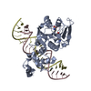
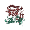
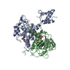
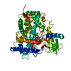
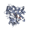
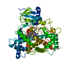
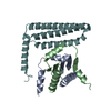
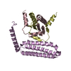
[English] 日本語
Yorodumi
- PDB-5f28: Crystal structure of FAT domain of Focal Adhesion Kinase (FAK) bo... -

+
Open data


ID or keywords:

Loading...

-
Basic information

Entry
Database: PDB / ID: 5f28
TitleCrystal structure of FAT domain of Focal Adhesion Kinase (FAK) bound to the transcription factor MEF2C
Components
  • Focal adhesion kinase 1PTK2
  • MEF2C
KeywordsTranscription / protein binding / Transcription Factor / Kinase / Cardiovascular disease
Function / homology
Function and homology information


primary heart field specification / sinoatrial valve morphogenesis / positive regulation of alkaline phosphatase activity / nephron tubule epithelial cell differentiation / muscle cell fate determination / cartilage morphogenesis / regulation of sarcomere organization / cardiac ventricle formation / glomerulus morphogenesis / epithelial cell proliferation involved in renal tubule morphogenesis ...primary heart field specification / sinoatrial valve morphogenesis / positive regulation of alkaline phosphatase activity / nephron tubule epithelial cell differentiation / muscle cell fate determination / cartilage morphogenesis / regulation of sarcomere organization / cardiac ventricle formation / glomerulus morphogenesis / epithelial cell proliferation involved in renal tubule morphogenesis / positive regulation of skeletal muscle cell differentiation / DCC mediated attractive signaling / positive regulation of macrophage apoptotic process / Apoptotic cleavage of cellular proteins / DNA-binding transcription factor binding => GO:0140297 / MET activates PTK2 signaling / transdifferentiation / HMG box domain binding / NCAM signaling for neurite out-growth / regulation of synaptic activity / secondary heart field specification / cellular response to xenobiotic stimulus => GO:0071466 / negative regulation of synapse assembly / renal tubule morphogenesis / cellular response to trichostatin A / cell morphogenesis involved in neuron differentiation / EPHB-mediated forward signaling / central nervous system neuron axonogenesis / ventricular cardiac muscle cell differentiation / positive regulation of behavioral fear response / embryonic viscerocranium morphogenesis / RHO GTPases Activate WASPs and WAVEs / regulation of substrate adhesion-dependent cell spreading / positive regulation of cell proliferation in bone marrow / GRB2:SOS provides linkage to MAPK signaling for Integrins / positive regulation of cardiac muscle cell differentiation / Integrin signaling / p130Cas linkage to MAPK signaling for integrins / RAF/MAP kinase cascade / Extra-nuclear estrogen signaling / regulation of dendritic spine development / intracellular chloride ion homeostasis / embryonic skeletal system morphogenesis / negative regulation of axonogenesis / regulation of epithelial cell migration / regulation of neurotransmitter secretion / Regulation of actin dynamics for phagocytic cup formation / negative regulation of organ growth / negative regulation of vascular associated smooth muscle cell migration / regulation of megakaryocyte differentiation / positive regulation of skeletal muscle tissue development / regulation of germinal center formation / smooth muscle cell differentiation / melanocyte differentiation / regulation of modification of postsynaptic actin cytoskeleton / cardiac muscle cell differentiation / germinal center formation / Myogenesis / regulation of AMPA receptor activity / endochondral ossification / positive regulation of ubiquitin-dependent protein catabolic process / JUN kinase binding / cellular response to fluid shear stress / VEGFA-VEGFR2 Pathway / cardiac muscle hypertrophy in response to stress / signal complex assembly / negative regulation of ossification / positive regulation of macrophage proliferation / neural crest cell differentiation / positive regulation of fibroblast migration / nuclear migration / cellular response to parathyroid hormone stimulus / negative regulation of vascular endothelial cell proliferation / regulation of NMDA receptor activity / growth hormone receptor signaling pathway / platelet formation / regulation of osteoblast differentiation / regulation of synapse assembly / regulation of focal adhesion assembly / organ growth / positive regulation of wound healing / positive regulation of macrophage chemotaxis / positive regulation of epithelial cell migration / roof of mouth development / negative regulation of cell-cell adhesion / positive regulation of cardiac muscle hypertrophy / heart looping / positive regulation of smooth muscle cell migration / blood vessel development / B cell proliferation / outflow tract morphogenesis / skeletal muscle cell differentiation / minor groove of adenine-thymine-rich DNA binding / negative regulation of vascular associated smooth muscle cell proliferation / regulation of cell adhesion mediated by integrin / monocyte differentiation / B cell homeostasis / humoral immune response / negative regulation of blood vessel endothelial cell migration / sarcoplasm
Similarity search - Function
SRF-like / Transcription factor, MADS-box / Holliday junction regulator protein family C-terminal / Holliday junction regulator protein family C-terminal repeat / MADS MEF2-like / Transcription factor, MADS-box / Transcription factor, MADS-box superfamily / SRF-type transcription factor (DNA-binding and dimerisation domain) / MADS-box domain signature. / MADS-box domain profile. ...SRF-like / Transcription factor, MADS-box / Holliday junction regulator protein family C-terminal / Holliday junction regulator protein family C-terminal repeat / MADS MEF2-like / Transcription factor, MADS-box / Transcription factor, MADS-box superfamily / SRF-type transcription factor (DNA-binding and dimerisation domain) / MADS-box domain signature. / MADS-box domain profile. / MADS / Single helix bin / Nucleotidyltransferases domain 2 / Focal adhesion kinase, targeting (FAT) domain / Focal adhesion kinase, targeting (FAT) domain superfamily / Focal adhesion kinase, N-terminal / FAK1/PYK2, FERM domain C-lobe / Focal adhesion targeting region / FERM N-terminal domain / : / FAK1/PYK2, FERM domain C-lobe / FERM domain signature 2. / FERM central domain / FERM/acyl-CoA-binding protein superfamily / FERM central domain / FERM superfamily, second domain / FERM domain / FERM domain profile. / Band 4.1 domain / Band 4.1 homologues / Four Helix Bundle (Hemerythrin (Met), subunit A) / Single alpha-helices involved in coiled-coils or other helix-helix interfaces / Tyrosine-protein kinase, catalytic domain / Tyrosine kinase, catalytic domain / Tyrosine protein kinases specific active-site signature. / PH-like domain superfamily / Tyrosine-protein kinase, active site / Protein tyrosine and serine/threonine kinase / Serine-threonine/tyrosine-protein kinase, catalytic domain / Ubiquitin-like domain superfamily / Protein kinase, ATP binding site / Protein kinases ATP-binding region signature. / Protein kinase domain profile. / Protein kinase domain / Protein kinase-like domain superfamily / Up-down Bundle / 3-Layer(aba) Sandwich / Mainly Alpha / Alpha Beta
Similarity search - Domain/homology
Focal adhesion kinase 1 / Myocyte-specific enhancer factor 2C
Similarity search - Component
Biological speciesMus musculus (house mouse)
MethodX-RAY DIFFRACTION / SYNCHROTRON / MOLECULAR REPLACEMENT / Resolution: 2.9 Å
AuthorsCardoso, A.C. / Ambrosio, A.L.B. / Dessen, A. / Franchini, K.G.
Funding support Brazil, 1items
OrganizationGrant numberCountry
Sao Paulo Research Foundation (FAPESP)2008/53519-5 Brazil
CitationJournal: Structure / Year: 2016
Title: FAK Forms a Complex with MEF2 to Couple Biomechanical Signaling to Transcription in Cardiomyocytes.
Authors: Cardoso, A.C. / Pereira, A.H.M. / Ambrosio, A.L.B. / Consonni, S.R. / Rocha de Oliveira, R. / Bajgelman, M.C. / Dias, S.M.G. / Franchini, K.G.
History
DepositionDec 1, 2015Deposition site: RCSB / Processing site: RCSB
Revision 1.0Jul 13, 2016Provider: repository / Type: Initial release
Revision 1.1Jan 23, 2019Group: Advisory / Data collection ...Advisory / Data collection / Database references / Derived calculations
Category: citation / citation_author ...citation / citation_author / pdbx_struct_oper_list / pdbx_unobs_or_zero_occ_atoms
Item: _citation.country / _citation.journal_abbrev ..._citation.country / _citation.journal_abbrev / _citation.journal_id_ASTM / _citation.journal_id_CSD / _citation.journal_id_ISSN / _citation.journal_volume / _citation.page_first / _citation.page_last / _citation.pdbx_database_id_DOI / _citation.pdbx_database_id_PubMed / _citation.title / _citation.year / _pdbx_struct_oper_list.symmetry_operation
Revision 1.2Apr 17, 2019Group: Author supporting evidence / Data collection / Category: pdbx_audit_support / Item: _pdbx_audit_support.funding_organization
Revision 1.3Jan 1, 2020Group: Author supporting evidence / Category: pdbx_audit_support / Item: _pdbx_audit_support.funding_organization
Revision 1.4Sep 27, 2023Group: Advisory / Data collection ...Advisory / Data collection / Database references / Refinement description
Category: chem_comp_atom / chem_comp_bond ...chem_comp_atom / chem_comp_bond / database_2 / pdbx_initial_refinement_model / pdbx_unobs_or_zero_occ_atoms
Item: _database_2.pdbx_DOI / _database_2.pdbx_database_accession

-
Structure visualization

Structure viewerMolecule:
MolmilJmol/JSmol

Downloads & links

-
Assembly

Deposited unit
A: MEF2C
B: MEF2C
C: MEF2C
D: MEF2C
E: Focal adhesion kinase 1
F: Focal adhesion kinase 1
G: Focal adhesion kinase 1


Theoretical massNumber of molelcules
Total (without water)94,4057
Polymers94,4057
Non-polymers00
Water1,04558
1
A: MEF2C
B: MEF2C
F: Focal adhesion kinase 1


Theoretical massNumber of molelcules
Total (without water)38,9373
Polymers38,9373
Non-polymers00
Water543
TypeNameSymmetry operationNumber
identity operation1_555x,y,z1
2
C: MEF2C
D: MEF2C
E: Focal adhesion kinase 1


Theoretical massNumber of molelcules
Total (without water)38,9373
Polymers38,9373
Non-polymers00
Water543
TypeNameSymmetry operationNumber
identity operation1_555x,y,z1
3
A: MEF2C
B: MEF2C
C: MEF2C
D: MEF2C
E: Focal adhesion kinase 1
F: Focal adhesion kinase 1
G: Focal adhesion kinase 1

A: MEF2C
B: MEF2C
C: MEF2C
D: MEF2C
E: Focal adhesion kinase 1
F: Focal adhesion kinase 1
G: Focal adhesion kinase 1


Theoretical massNumber of molelcules
Total (without water)188,81114
Polymers188,81114
Non-polymers00
Water25214
TypeNameSymmetry operationNumber
identity operation1_555x,y,z1
crystal symmetry operation7_555y,x,-z1
Buried area42370 Å2
ΔGint-317 kcal/mol
Surface area65140 Å2
MethodPISA
Unit cell
Length a, b, c (Å)139.210, 139.210, 90.350
Angle α, β, γ (deg.)90.00, 90.00, 90.00
Int Tables number94
Space group name H-MP42212

-
Components

#1: Protein
MEF2C


Mass: 11202.983 Da / Num. of mol.: 4 / Fragment: UNP residues 1-95
Source method: isolated from a genetically manipulated source
Source: (gene. exp.) Mus musculus (house mouse) / Plasmid: pETSUMO / Details (production host): Home-made / Production host: Escherichia coli (E. coli) / Strain (production host): C41 (DE3) / References: UniProt: Q8CFN5
#2: Protein Focal adhesion kinase 1 / PTK2


Mass: 16531.158 Da / Num. of mol.: 3 / Fragment: UNP residues 935-1083
Source method: isolated from a genetically manipulated source
Source: (gene. exp.) Mus musculus (house mouse) / Plasmid: pET28a-TEV / Production host: Escherichia coli (E. coli) / Strain (production host): C41 (DE3) / References: UniProt: P34152
#3: Water ChemComp-HOH / water / Water


Mass: 18.015 Da / Num. of mol.: 58 / Source method: isolated from a natural source / Formula: H2O

-
Experimental details

-
Experiment

ExperimentMethod: X-RAY DIFFRACTION

-
Sample preparation

CrystalDensity Matthews: 2.31 Å3/Da / Density % sol: 46.7 %
Crystal growTemperature: 291 K / Method: vapor diffusion, hanging drop / pH: 6.5
Details: 0.1M Magnseium acetate, 0.1M MES, pH 6.5, 12% PEG 8000

-
Data collection

DiffractionMean temperature: 100 K
Diffraction sourceSource: SYNCHROTRON / Site: ESRF / Beamline: ID23-2 / Wavelength: 0.8729 Å
DetectorType: MARMOSAIC 225 mm CCD / Detector: CCD / Date: Apr 11, 2013
RadiationMonochromator: Silicon 111 crystal / Protocol: SINGLE WAVELENGTH / Monochromatic (M) / Laue (L): M / Scattering type: x-ray
Radiation wavelengthWavelength: 0.8729 Å / Relative weight: 1
ReflectionResolution: 2.9→49.22 Å / Num. obs: 37566 / % possible obs: 100 % / Redundancy: 13.7 % / Rmerge(I) obs: 0.344 / Net I/σ(I): 8.7
Reflection shellResolution: 2.9→3.08 Å / Redundancy: 13.7 % / Rmerge(I) obs: 1.559 / Mean I/σ(I) obs: 1.9 / % possible all: 100

-
Processing

Software
NameVersionClassification
PHENIX(dev_2196)refinement
MOSFLMdata reduction
Aimlessdata scaling
PHASERphasing
RefinementMethod to determine structure: MOLECULAR REPLACEMENT
Starting model: PDB entries 1K40 (FAT) and 3KOV (MEF2)
Resolution: 2.9→43.221 Å / SU ML: 0.42 / Cross valid method: FREE R-VALUE / σ(F): 0.34 / Phase error: 25.04 / Stereochemistry target values: ML
RfactorNum. reflection% reflectionSelection details
Rfree0.2354 1880 5 %Random selection
Rwork0.2084 ---
obs0.2097 37566 99.96 %-
Solvent computationShrinkage radii: 0.9 Å / VDW probe radii: 1.11 Å / Solvent model: FLAT BULK SOLVENT MODEL
Refinement stepCycle: LAST / Resolution: 2.9→43.221 Å
ProteinNucleic acidLigandSolventTotal
Num. atoms5360 0 0 58 5418
Refine LS restraints
Refine-IDTypeDev idealNumber
X-RAY DIFFRACTIONf_bond_d0.0045429
X-RAY DIFFRACTIONf_angle_d0.5937318
X-RAY DIFFRACTIONf_dihedral_angle_d16.1382095
X-RAY DIFFRACTIONf_chiral_restr0.038873
X-RAY DIFFRACTIONf_plane_restr0.004919
LS refinement shell
Resolution (Å)Rfactor RfreeNum. reflection RfreeRfactor RworkNum. reflection RworkRefine-ID% reflection obs (%)
2.9-2.97840.37621430.36162714X-RAY DIFFRACTION100
2.9784-3.06610.38341440.32382747X-RAY DIFFRACTION100
3.0661-3.1650.31261290.29732754X-RAY DIFFRACTION100
3.165-3.27810.32411440.27932758X-RAY DIFFRACTION100
3.2781-3.40930.27151430.26362751X-RAY DIFFRACTION100
3.4093-3.56440.25551480.23882761X-RAY DIFFRACTION100
3.5644-3.75220.26411740.20792721X-RAY DIFFRACTION100
3.7522-3.98710.24381410.192738X-RAY DIFFRACTION100
3.9871-4.29470.18571480.16522742X-RAY DIFFRACTION100
4.2947-4.72640.20471200.15632768X-RAY DIFFRACTION100
4.7264-5.40920.18321410.15152753X-RAY DIFFRACTION100
5.4092-6.81080.22311600.20962732X-RAY DIFFRACTION100
6.8108-43.2260.14711450.16072747X-RAY DIFFRACTION100

+
About Yorodumi

-
News

-
Feb 9, 2022. New format data for meta-information of EMDB entries

New format data for meta-information of EMDB entries

  • Version 3 of the EMDB header file is now the official format.
  • The previous official version 1.9 will be removed from the archive.

Related info.:EMDB header

External links:wwPDB to switch to version 3 of the EMDB data model

-
Aug 12, 2020. Covid-19 info

Covid-19 info

URL: https://pdbj.org/emnavi/covid19.php

New page: Covid-19 featured information page in EM Navigator.

Related info.:Covid-19 info / Mar 5, 2020. Novel coronavirus structure data

+
Mar 5, 2020. Novel coronavirus structure data

Novel coronavirus structure data

Related info.:Yorodumi Speices / Aug 12, 2020. Covid-19 info

External links:COVID-19 featured content - PDBj / Molecule of the Month (242):Coronavirus Proteases

+
Jan 31, 2019. EMDB accession codes are about to change! (news from PDBe EMDB page)

EMDB accession codes are about to change! (news from PDBe EMDB page)

  • The allocation of 4 digits for EMDB accession codes will soon come to an end. Whilst these codes will remain in use, new EMDB accession codes will include an additional digit and will expand incrementally as the available range of codes is exhausted. The current 4-digit format prefixed with “EMD-” (i.e. EMD-XXXX) will advance to a 5-digit format (i.e. EMD-XXXXX), and so on. It is currently estimated that the 4-digit codes will be depleted around Spring 2019, at which point the 5-digit format will come into force.
  • The EM Navigator/Yorodumi systems omit the EMD- prefix.

Related info.:Q: What is EMD? / ID/Accession-code notation in Yorodumi/EM Navigator

External links:EMDB Accession Codes are Changing Soon! / Contact to PDBj

+
Jul 12, 2017. Major update of PDB

Major update of PDB

  • wwPDB released updated PDB data conforming to the new PDBx/mmCIF dictionary.
  • This is a major update changing the version number from 4 to 5, and with Remediation, in which all the entries are updated.
  • In this update, many items about electron microscopy experimental information are reorganized (e.g. em_software).
  • Now, EM Navigator and Yorodumi are based on the updated data.

External links:wwPDB Remediation / Enriched Model Files Conforming to OneDep Data Standards Now Available in the PDB FTP Archive

-
Yorodumi

Thousand views of thousand structures

  • Yorodumi is a browser for structure data from EMDB, PDB, SASBDB, etc.
  • This page is also the successor to EM Navigator detail page, and also detail information page/front-end page for Omokage search.
  • The word "yorodu" (or yorozu) is an old Japanese word meaning "ten thousand". "mi" (miru) is to see.

Related info.:EMDB / PDB / SASBDB / Comparison of 3 databanks / Yorodumi Search / Aug 31, 2016. New EM Navigator & Yorodumi / Yorodumi Papers / Jmol/JSmol / Function and homology information / Changes in new EM Navigator and Yorodumi

Read more