 Movie
Movie Controller

[English] 日本語
 Yorodumi
- PDB-5f1b: Structural basis of Ebola virus entry: viral glycoprotein bound t... -
+ Open data

- Basic information
| Entry | Database: PDB / ID: 5f1b | |||||||||
|---|---|---|---|---|---|---|---|---|---|---|
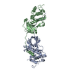
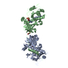
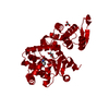
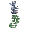
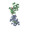
| Title | Structural basis of Ebola virus entry: viral glycoprotein bound to its endosomal receptor Niemann-Pick C1 | |||||||||
 Components Components |
| |||||||||
 Keywords Keywords |  VIRAL PROTEIN/TRANSPORT PROTEIN / Ebola virus / glycoprotein / NPC1-C / VIRAL PROTEIN-TRANSPORT PROTEIN complex VIRAL PROTEIN/TRANSPORT PROTEIN / Ebola virus / glycoprotein / NPC1-C / VIRAL PROTEIN-TRANSPORT PROTEIN complex | |||||||||
| Function / homology |  Function and homology information Function and homology informationcyclodextrin metabolic process / cholesterol storage / membrane raft organization / : / intracellular cholesterol transport / intracellular lipid transport / intestinal cholesterol absorption / LDL clearance / programmed cell death / negative regulation of epithelial cell apoptotic process ...cyclodextrin metabolic process / cholesterol storage / membrane raft organization / : / intracellular cholesterol transport / intracellular lipid transport / intestinal cholesterol absorption / LDL clearance / programmed cell death / negative regulation of epithelial cell apoptotic process / cholesterol transport / bile acid metabolic process / establishment of protein localization to membrane / cholesterol efflux / adult walking behavior / lysosomal transport / cholesterol binding / negative regulation of macroautophagy / cellular response to steroid hormone stimulus / protein glycosylation / cellular response to low-density lipoprotein particle stimulus / response to cadmium ion / negative regulation of TORC1 signaling / cholesterol metabolic process / neurogenesis / cholesterol homeostasis / liver development / macroautophagy / autophagy / endocytosis / transmembrane signaling receptor activity / virus receptor activity / signaling receptor activity / nuclear envelope / late endosome membrane / gene expression / clathrin-dependent endocytosis of virus by host cell / suppression by virus of host tetherin activity / entry receptor-mediated virion attachment to host cell / lysosome / response to xenobiotic stimulus / symbiont entry into host cell / membrane raft / lysosomal membrane / fusion of virus membrane with host endosome membrane / viral envelope / lipid binding / symbiont-mediated suppression of host type I interferon-mediated signaling pathway / host cell plasma membrane / virion membrane / perinuclear region of cytoplasm / Golgi apparatus / endoplasmic reticulum / extracellular exosome / extracellular region / membrane / plasma membraneSimilarity search - Function | |||||||||
| Biological species |   Zaire ebolavirus Zaire ebolavirus Homo sapiens (human) Homo sapiens (human) | |||||||||
| Method | X-RAY DIFFRACTION / SYNCHROTRON / MOLECULAR REPLACEMENT / Resolution: 2.3 Å | |||||||||
 Authors Authors | Wang, H. / Shi, Y. / Song, J. / Qi, J. / Lu, G. / Yan, J. / Gao, G.F. | |||||||||
| Funding support |  China, 1items China, 1items
| |||||||||
 Citation Citation | Journal: Cell / Year: 2016 Title: Ebola Viral Glycoprotein Bound to Its Endosomal Receptor Niemann-Pick C1. Authors: Wang, H. / Shi, Y. / Song, J. / Qi, J. / Lu, G. / Yan, J. / Gao, G.F. | |||||||||
| History |
|
- Structure visualization
| Structure viewer | Molecule: MolmilJmol/JSmol |
|---|
- Downloads & links
-Download
| PDBx/mmCIF format | 5f1b.cif.gz | 205.5 KB | Display | PDBx/mmCIF format |
|---|---|---|---|---|
| PDB format | pdb5f1b.ent.gz | 162.1 KB | Display | PDB format |
| PDBx/mmJSON format | 5f1b.json.gz | Tree view | PDBx/mmJSON format | |
| Others |  Other downloads Other downloads |
-Validation report
| Arichive directory | https://data.pdbj.org/pub/pdb/validation_reports/f1/5f1bftp://data.pdbj.org/pub/pdb/validation_reports/f1/5f1b | HTTPS FTP |
|---|
-Related structure data
| Related structure data |  5f18C 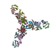 3csyS C: citing same article ( S: Starting model for refinement |
|---|---|
| Similar structure data |


-Links
 PDBj
PDBj


- Assembly
| Deposited unit | 
| ||||||||
|---|---|---|---|---|---|---|---|---|---|
| 1 |
| ||||||||
| Unit cell |
|
-Components
| #1: Protein | Mass: 17194.543 Da / Num. of mol.: 1 / Fragment: UNP RESIDUES 32-188 Source method: isolated from a genetically manipulated source Source: (gene. exp.) Zaire ebolavirus / Strain: Kikwit-95 / Gene: GP / Cell line (production host): High Five cells / Production host:  Trichoplusia ni (cabbage looper) / References: UniProt: P87666 Trichoplusia ni (cabbage looper) / References: UniProt: P87666 |
|---|---|
| #2: Protein | Mass: 14882.807 Da / Num. of mol.: 1 / Fragment: UNP RESIDUES 509-598 Source method: isolated from a genetically manipulated source Source: (gene. exp.) Zaire ebolavirus / Strain: Kikwit-95 / Gene: GP / Cell line (production host): High Five cells / Production host: Trichoplusia ni (cabbage looper) / References: UniProt: P87666 |
| #3: Protein | Mass: 29436.582 Da / Num. of mol.: 1 / Fragment: UNP RESIDUES 374-620 Source method: isolated from a genetically manipulated source Source: (gene. exp.) Homo sapiens (human) / Gene: NPC1 / Production host:  Escherichia coli (E. coli) / References: UniProt: O15118 Escherichia coli (E. coli) / References: UniProt: O15118 |
| #4: Polysaccharide | 2-acetamido-2-deoxy-beta-D-glucopyranose-(1-4)-2-acetamido-2-deoxy-beta-D-glucopyranose / Mass: 424.401 Da / Num. of mol.: 1 Source method: isolated from a genetically manipulated source |
| #5: Water | ChemComp-HOH / Water Mass: 18.015 Da / Num. of mol.: 171 / Source method: isolated from a natural source / Formula: H2O Mass: 18.015 Da / Num. of mol.: 171 / Source method: isolated from a natural source / Formula: H2O |
-Experimental details
-Experiment
| Experiment | Method: X-RAY DIFFRACTION |
|---|
- Sample preparation
| Crystal | Density Matthews: 2.53 Å3/Da / Density % sol: 51.41 % |
|---|---|
| Crystal grow | Temperature: 291 K / Method: vapor diffusion, sitting drop / pH: 5.5 Details: 15% (v/v) pentaerythritolpropoxylate, 0.2M sodium chloride, pH 5.5, 0.1M MES-NaOH |
-Data collection
| Diffraction | Mean temperature: 100 K |
|---|---|
| Diffraction source | Source: SYNCHROTRON / Site: SSRF / Beamline: BL19U1 / Wavelength: 0.9793 Å |
| Detector | Type: PSI PILATUS 6M / Detector: PIXEL / Date: Jun 18, 2015 |
| Radiation | Protocol: SINGLE WAVELENGTH / Monochromatic (M) / Laue (L): M / Scattering type: x-ray |
| Radiation wavelength | Wavelength: 0.9793 Å / Relative weight: 1 |
| Reflection | Resolution: 2.3→50 Å / Num. obs: 27329 / % possible obs: 100 % / Redundancy: 19.6 % / Net I/σ(I): 32.6 |
- Processing
| Software |
| |||||||||||||||||||||||||||||||||||||||||||||||||||||||||||||||||||||||||||||
|---|---|---|---|---|---|---|---|---|---|---|---|---|---|---|---|---|---|---|---|---|---|---|---|---|---|---|---|---|---|---|---|---|---|---|---|---|---|---|---|---|---|---|---|---|---|---|---|---|---|---|---|---|---|---|---|---|---|---|---|---|---|---|---|---|---|---|---|---|---|---|---|---|---|---|---|---|---|---|
| Refinement | Method to determine structure: MOLECULAR REPLACEMENT Starting model: 3CSY Resolution: 2.3→41.858 Å / SU ML: 0.28 / Cross valid method: FREE R-VALUE / σ(F): 1.36 / Phase error: 23.61 / Stereochemistry target values: ML
| |||||||||||||||||||||||||||||||||||||||||||||||||||||||||||||||||||||||||||||
| Solvent computation | Shrinkage radii: 0.9 Å / VDW probe radii: 1.11 Å / Solvent model: FLAT BULK SOLVENT MODEL | |||||||||||||||||||||||||||||||||||||||||||||||||||||||||||||||||||||||||||||
| Refinement step | Cycle: LAST / Resolution: 2.3→41.858 Å
| |||||||||||||||||||||||||||||||||||||||||||||||||||||||||||||||||||||||||||||
| Refine LS restraints |
| |||||||||||||||||||||||||||||||||||||||||||||||||||||||||||||||||||||||||||||
| LS refinement shell |
| |||||||||||||||||||||||||||||||||||||||||||||||||||||||||||||||||||||||||||||
| Refinement TLS params. | Method: refined / Origin x: 30.2833 Å / Origin y: -16.3893 Å / Origin z: -320.9409 Å
| |||||||||||||||||||||||||||||||||||||||||||||||||||||||||||||||||||||||||||||
| Refinement TLS group | Selection details: all |
