 Movie
Movie Controller

[English] 日本語
 Yorodumi
- PDB-5ezf: Racemic crystal structures of Pribnow box consensus promoter sequ... -
+ Open data

- Basic information
| Entry | Database: PDB / ID: 5ezf | ||||||
|---|---|---|---|---|---|---|---|
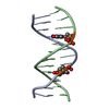
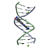
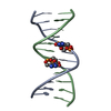
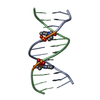
| Title | Racemic crystal structures of Pribnow box consensus promoter sequence (Pbca) | ||||||
 Components Components |
| ||||||
 Keywords Keywords |  DNA / Pribnow box consensus sequence / -10 element / transcription initiation / B-DNA double helix / DNA double helix DNA / Pribnow box consensus sequence / -10 element / transcription initiation / B-DNA double helix / DNA double helix | ||||||
| Function / homology | DNA / DNA (> 10) Function and homology information Function and homology information | ||||||
| Biological species | synthetic construct (others) | ||||||
| Method | X-RAY DIFFRACTION / SYNCHROTRON / MOLECULAR REPLACEMENT / Resolution: 1.65 Å | ||||||
 Authors Authors | Mandal, P.K. / Collie, G.W. / Kauffmann, B. / Srivastava, S.C. / Huc, I. | ||||||
| Funding support |  France, 1items France, 1items
| ||||||
 Citation Citation | Journal: Nucleic Acids Res. / Year: 2016 Title: Structure elucidation of the Pribnow box consensus promoter sequence by racemic DNA crystallography. Authors: Mandal, P.K. / Collie, G.W. / Srivastava, S.C. / Kauffmann, B. / Huc, I. #1: Journal: Angew. Chem. Int. Ed. / Year: 2014Title: Racemic DNA Crystallography Authors: Mandal, P.K. / Collie, G.W. / Kauffmann, B. / Huc, I. | ||||||
| History |
|
- Structure visualization
| Structure viewer | Molecule: MolmilJmol/JSmol |
|---|
- Downloads & links
-Download
| PDBx/mmCIF format | 5ezf.cif.gz | 39.1 KB | Display | PDBx/mmCIF format |
|---|---|---|---|---|
| PDB format | pdb5ezf.ent.gz | 27 KB | Display | PDB format |
| PDBx/mmJSON format | 5ezf.json.gz | Tree view | PDBx/mmJSON format | |
| Others |  Other downloads Other downloads |
-Validation report
| Arichive directory | https://data.pdbj.org/pub/pdb/validation_reports/ez/5ezfftp://data.pdbj.org/pub/pdb/validation_reports/ez/5ezf | HTTPS FTP |
|---|
-Related structure data
| Related structure data |  5et9C  5ewbC  5eyqC  5f26C  5j0eC  1fq2S C: citing same article ( S: Starting model for refinement |
|---|---|
| Similar structure data |


-Links
 PDBj
PDBj


- Assembly
| Deposited unit | 
| ||||||||
|---|---|---|---|---|---|---|---|---|---|
| 1 |
| ||||||||
| Unit cell |
|
-Components
| #1: DNA chain | Mass: 3662.404 Da / Num. of mol.: 1 / Source method: obtained synthetically / Source: (synth.) synthetic construct (others) | ||
|---|---|---|---|
| #2: DNA chain | Mass: 3662.404 Da / Num. of mol.: 1 / Source method: obtained synthetically / Source: (synth.) synthetic construct (others) | ||
| #3: Chemical |   Mass: 40.078 Da / Num. of mol.: 2 / Source method: obtained synthetically / Formula: Ca Mass: 40.078 Da / Num. of mol.: 2 / Source method: obtained synthetically / Formula: Ca#4: Water | ChemComp-HOH / | Water Mass: 18.015 Da / Num. of mol.: 52 / Source method: isolated from a natural source / Formula: H2O Mass: 18.015 Da / Num. of mol.: 52 / Source method: isolated from a natural source / Formula: H2O |
-Experimental details
-Experiment
| Experiment | Method: X-RAY DIFFRACTION |
|---|
- Sample preparation
| Crystal | Density Matthews: 1.85 Å3/Da / Density % sol: 55 % |
|---|---|
| Crystal grow | Temperature: 293 K / Method: vapor diffusion, hanging drop / pH: 7.5 Details: Racemic DNA mixture*, HEPES, calcium chloride, PEG400 * For crystallization, we used four strands 1) d(CGCTATAATGCG) with L-sugars 2) d(CGCATTATAGCG) with L-sugars and 3) d(CGCTATAATGCG) ...Details: Racemic DNA mixture*, HEPES, calcium chloride, PEG400 * For crystallization, we used four strands 1) d(CGCTATAATGCG) with L-sugars 2) d(CGCATTATAGCG) with L-sugars and 3) d(CGCTATAATGCG) with D-sugars 4) d(CGCATTATAGCG) with D-sugars Enantio-pure DNA solutions were prepared first by de-naturation followed by slow cooling down process to ensure proper folding of the hetero-duplex formed between the non-self complementary strands. After slow-annealing, the enantiopure solutions were mixed in equimolar ratio and this racemic DNA mixture was used for crystallization. |
-Data collection
| Diffraction | Mean temperature: 100 K |
|---|---|
| Diffraction source | Source: SYNCHROTRON / Site: SOLEIL / Beamline: PROXIMA 1 / Wavelength: 0.9785 Å |
| Detector | Type: DECTRIS PILATUS 6M / Detector: PIXEL / Date: Jun 11, 2015 |
| Radiation | Protocol: SINGLE WAVELENGTH / Monochromatic (M) / Laue (L): M / Scattering type: x-ray |
| Radiation wavelength | Wavelength: 0.9785 Å / Relative weight: 1 |
| Reflection | Resolution: 1.65→32.86 Å / Num. obs: 12549 / % possible obs: 99.86 % / Redundancy: 1.9 % / Rmerge(I) obs: 0.019 / Net I/σ(I): 16.92 |
| Reflection shell | Resolution: 1.65→1.79 Å / Redundancy: 1.9 % / Rmerge(I) obs: 0.268 / Mean I/σ(I) obs: 2.75 / % possible all: 99.76 |
- Processing
| Software |
| |||||||||||||||||||||||||||||||||||||||||||||||||||||||||||||||||||||||||||
|---|---|---|---|---|---|---|---|---|---|---|---|---|---|---|---|---|---|---|---|---|---|---|---|---|---|---|---|---|---|---|---|---|---|---|---|---|---|---|---|---|---|---|---|---|---|---|---|---|---|---|---|---|---|---|---|---|---|---|---|---|---|---|---|---|---|---|---|---|---|---|---|---|---|---|---|---|
| Refinement | Method to determine structure: MOLECULAR REPLACEMENT Starting model: 1FQ2 Resolution: 1.65→32.856 Å / SU ML: 0.22 / Cross valid method: FREE R-VALUE / σ(F): 1.33 / Phase error: 31.68 / Stereochemistry target values: ML
| |||||||||||||||||||||||||||||||||||||||||||||||||||||||||||||||||||||||||||
| Solvent computation | Shrinkage radii: 0.9 Å / VDW probe radii: 1.11 Å / Solvent model: FLAT BULK SOLVENT MODEL | |||||||||||||||||||||||||||||||||||||||||||||||||||||||||||||||||||||||||||
| Refinement step | Cycle: LAST / Resolution: 1.65→32.856 Å
| |||||||||||||||||||||||||||||||||||||||||||||||||||||||||||||||||||||||||||
| Refine LS restraints |
| |||||||||||||||||||||||||||||||||||||||||||||||||||||||||||||||||||||||||||
| LS refinement shell |
| |||||||||||||||||||||||||||||||||||||||||||||||||||||||||||||||||||||||||||
| Refinement TLS params. | Method: refined / Refine-ID: X-RAY DIFFRACTION
| |||||||||||||||||||||||||||||||||||||||||||||||||||||||||||||||||||||||||||
| Refinement TLS group |
|
