 Movie
Movie Controller

[English] 日本語
 Yorodumi
- PDB-3a4k: Crystal structural analysis of HindIII restriction endonuclease i... -
+ Open data

- Basic information
| Entry | Database: PDB / ID: 3a4k | ||||||
|---|---|---|---|---|---|---|---|
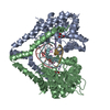
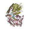
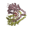
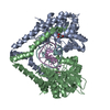
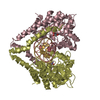
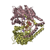
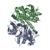
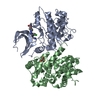
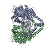
| Title | Crystal structural analysis of HindIII restriction endonuclease in complex with cognate DNA and divalent cations at 2.17 angstrom resolution | ||||||
 Components Components |
| ||||||
 Keywords Keywords | HYDROLASE/DNA / Type II restriction enzyme HindIII(E.C.3.1.21.4)/DNA / hydrolase-DNA COMPLEX / Endonuclease / Hydrolase / Nuclease / Restriction system | ||||||
| Function / homology |  Function and homology information Function and homology informationtype II site-specific deoxyribonuclease / type II site-specific deoxyribonuclease activity / DNA restriction-modification system Similarity search - Function | ||||||
| Biological species |  Haemophilus influenzae (bacteria) Haemophilus influenzae (bacteria) | ||||||
| Method |  X-RAY DIFFRACTION / SYNCHROTRON / MOLECULAR REPLACEMENT / Resolution: 2.17 Å X-RAY DIFFRACTION / SYNCHROTRON / MOLECULAR REPLACEMENT / Resolution: 2.17 Å | ||||||
 Authors Authors | Watanabe, N. / Sato, C. / Takasaki, Y. / Tanaka, I. | ||||||
 Citation Citation | Journal: Acta Crystallogr.,Sect.D / Year: 2009 Title: Structures of restriction endonuclease HindIII in complex with its cognate DNA and divalent cations Authors: Watanabe, N. / Takasaki, Y. / Sato, C. / Ando, S. / Tanaka, I. | ||||||
| History |
| ||||||
| Remark 650 | HELIX Determination method: Author determined |
- Structure visualization
| Structure viewer | Molecule: MolmilJmol/JSmol |
|---|
- Downloads & links
-Download
| PDBx/mmCIF format | 3a4k.cif.gz | 305.9 KB | Display | PDBx/mmCIF format |
|---|---|---|---|---|
| PDB format | pdb3a4k.ent.gz | 241.3 KB | Display | PDB format |
| PDBx/mmJSON format | 3a4k.json.gz | Tree view | PDBx/mmJSON format | |
| Others |  Other downloads Other downloads |
-Validation report
| Summary document | 3a4k_validation.pdf.gz | 527.3 KB | Display | wwPDB validaton report |
|---|---|---|---|---|
| Full document | 3a4k_full_validation.pdf.gz | 553.4 KB | Display | |
| Data in XML | 3a4k_validation.xml.gz | 51.7 KB | Display | |
| Data in CIF | 3a4k_validation.cif.gz | 74.5 KB | Display | |
| Arichive directory | https://data.pdbj.org/pub/pdb/validation_reports/a4/3a4kftp://data.pdbj.org/pub/pdb/validation_reports/a4/3a4k | HTTPS FTP |
-Related structure data
| Related structure data | 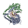 2e52SC S: Starting model for refinement C: citing same article ( |
|---|---|
| Similar structure data |

-Links
 PDBj
PDBj


- Assembly
| Deposited unit | 
| ||||||||
|---|---|---|---|---|---|---|---|---|---|
| 1 | 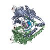
| ||||||||
| 2 | 
| ||||||||
| 3 | 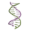
| ||||||||
| Unit cell |
|
-Components
-Protein , 1 types, 4 molecules ACBD
| #1: Protein | Mass: 35136.320 Da / Num. of mol.: 4 Source method: isolated from a genetically manipulated source Source: (gene. exp.) Haemophilus influenzae (bacteria) / Strain: Rd KW20 / Gene: hindIIIR / Plasmid: pET16b / Production host: References: UniProt: P43870, type II site-specific deoxyribonuclease |
|---|
-DNA chain , 3 types, 10 molecules EGIKFHJLMN
| #2: DNA chain | Mass: 1175.819 Da / Num. of mol.: 4 / Source method: obtained synthetically / Details: 5'-fragment of cleaved cognate DNA with HindIIIR #3: DNA chain | Mass: 2442.616 Da / Num. of mol.: 4 / Source method: obtained synthetically / Details: 3'-fragment of cleaved cognate DNA with HindIIIR #4: DNA chain | Mass: 3663.392 Da / Num. of mol.: 2 / Source method: obtained synthetically Details: This DNA sequence synthesized chemically contains cognate HindIIIR recognition sequence and elongated scaffolds on both sides |
|---|
-Non-polymers , 5 types, 587 molecules 


| #5: Chemical | ChemComp-MG /  Mass: 24.305 Da / Num. of mol.: 4 / Source method: obtained synthetically / Formula: Mg Mass: 24.305 Da / Num. of mol.: 4 / Source method: obtained synthetically / Formula: Mg#6: Chemical | ChemComp-MN /  Mass: 54.938 Da / Num. of mol.: 4 / Source method: obtained synthetically / Formula: Mn Mass: 54.938 Da / Num. of mol.: 4 / Source method: obtained synthetically / Formula: Mn#7: Chemical |  Mass: 92.094 Da / Num. of mol.: 2 / Source method: obtained synthetically / Formula: C3H8O3 Mass: 92.094 Da / Num. of mol.: 2 / Source method: obtained synthetically / Formula: C3H8O3#8: Chemical | ChemComp-ACT /  Mass: 59.044 Da / Num. of mol.: 4 / Source method: obtained synthetically / Formula: C2H3O2 Mass: 59.044 Da / Num. of mol.: 4 / Source method: obtained synthetically / Formula: C2H3O2#9: Water | ChemComp-HOH / | Mass: 18.015 Da / Num. of mol.: 573 / Source method: isolated from a natural source / Formula: H2O |
|---|
-Details
| Nonpolymer details | MG ION WAS USED IN THE EXPERIMENT, BUT METAL IONS AT SITES O, P, Q AND R IN THE STRUCTURE HAVE BEEN ...MG ION WAS USED IN THE EXPERIMENT |
|---|
-Experimental details
-Experiment
| Experiment | Method: X-RAY DIFFRACTION / Number of used crystals: 1 |
|---|
- Sample preparation
| Crystal | Density Matthews: 2.99 Å3/Da / Density % sol: 58.92 % |
|---|---|
| Crystal grow | Temperature: 293 K / Method: vapor diffusion, hanging drop / pH: 8 Details: PEG3350, ammonium acetate, Magnesium ion, glycerol, pH 8.0, VAPOR DIFFUSION, HANGING DROP, temperature 293K |
-Data collection
| Diffraction | Mean temperature: 100 K |
|---|---|
| Diffraction source | Source: SYNCHROTRON / Site: Photon Factory  / Beamline: BL-6A / Wavelength: 1 Å / Beamline: BL-6A / Wavelength: 1 Å |
| Detector | Type: ADSC QUANTUM 4r / Detector: CCD / Date: Oct 20, 2006 / Details: mirror |
| Radiation | Monochromator: Si(111) / Protocol: SINGLE WAVELENGTH / Monochromatic (M) / Laue (L): M / Scattering type: x-ray |
| Radiation wavelength | Wavelength: 1 Å / Relative weight: 1 |
| Reflection | Resolution: 2.17→50 Å / Num. obs: 100664 / % possible obs: 99.8 % / Redundancy: 1.9 % / Rmerge(I) obs: 0.063 / Net I/σ(I): 24.7 |
| Reflection shell | Resolution: 2.17→2.23 Å / Rmerge(I) obs: 0.404 / Mean I/σ(I) obs: 3.5 / % possible all: 98 |
- Processing
| Software |
| |||||||||||||||||||||||||||||||||||||||||||||||||||||||||||||||||
|---|---|---|---|---|---|---|---|---|---|---|---|---|---|---|---|---|---|---|---|---|---|---|---|---|---|---|---|---|---|---|---|---|---|---|---|---|---|---|---|---|---|---|---|---|---|---|---|---|---|---|---|---|---|---|---|---|---|---|---|---|---|---|---|---|---|---|
| Refinement | Method to determine structure: MOLECULAR REPLACEMENT Starting model: PDB ENTRY 2E52 Resolution: 2.17→41.67 Å / Cor.coef. Fo:Fc: 0.96 / Cor.coef. Fo:Fc free: 0.939 / Occupancy max: 1 / Occupancy min: 0.5 / SU B: 4.791 / SU ML: 0.125 / Isotropic thermal model: Isotropic / Cross valid method: THROUGHOUT / σ(F): 0 / ESU R: 0.195 / ESU R Free: 0.176 / Stereochemistry target values: MAXIMUM LIKELIHOOD
| |||||||||||||||||||||||||||||||||||||||||||||||||||||||||||||||||
| Solvent computation | Ion probe radii: 0.8 Å / Shrinkage radii: 0.8 Å / VDW probe radii: 1.2 Å / Solvent model: MASK | |||||||||||||||||||||||||||||||||||||||||||||||||||||||||||||||||
| Displacement parameters | Biso max: 85.53 Å2 / Biso mean: 36.106 Å2 / Biso min: 15.15 Å2
| |||||||||||||||||||||||||||||||||||||||||||||||||||||||||||||||||
| Refine analyze | Luzzati coordinate error obs: 0.2388 Å / Luzzati sigma a obs: 0.192792 Å | |||||||||||||||||||||||||||||||||||||||||||||||||||||||||||||||||
| Refinement step | Cycle: LAST / Resolution: 2.17→41.67 Å
| |||||||||||||||||||||||||||||||||||||||||||||||||||||||||||||||||
| Refine LS restraints |
| |||||||||||||||||||||||||||||||||||||||||||||||||||||||||||||||||
| LS refinement shell | Resolution: 2.169→2.226 Å / Total num. of bins used: 20
|
