 Movie
Movie Controller


+ Open data

- Basic information
| Entry | Database: PDB / ID: 5exa | ||||||
|---|---|---|---|---|---|---|---|
| Title | Small-molecule stabilization of the 14-3-3/Gab2 PPI interface | ||||||
 Components Components |
| ||||||
 Keywords Keywords |  SIGNALING PROTEIN / 14-3-3 / GAB2 / Protein / Diphosphorylation SIGNALING PROTEIN / 14-3-3 / GAB2 / Protein / Diphosphorylation | ||||||
| Function / homology |  Function and homology information Function and homology informationGolgi reassembly / regulation of synapse maturation / NOTCH4 Activation and Transmission of Signal to the Nucleus / establishment of Golgi localization / positive regulation of mast cell degranulation / STAT5 Activation / Rap1 signalling / phosphatidylinositol-3,4-bisphosphate binding / transmembrane receptor protein tyrosine kinase adaptor activity / Signaling by cytosolic FGFR1 fusion mutants ...Golgi reassembly / regulation of synapse maturation / NOTCH4 Activation and Transmission of Signal to the Nucleus / establishment of Golgi localization / positive regulation of mast cell degranulation / STAT5 Activation / Rap1 signalling / phosphatidylinositol-3,4-bisphosphate binding / transmembrane receptor protein tyrosine kinase adaptor activity / Signaling by cytosolic FGFR1 fusion mutants / Interleukin-15 signaling / negative regulation of protein localization to nucleus / KSRP (KHSRP) binds and destabilizes mRNA / GP1b-IX-V activation signalling / STAT5 activation downstream of FLT3 ITD mutants / phosphatidylinositol-3,4,5-trisphosphate binding / Regulation of localization of FOXO transcription factors / RET signaling / Interleukin-3, Interleukin-5 and GM-CSF signaling / PI3K Cascade / phosphoserine residue binding / Activation of BAD and translocation to mitochondria / Role of LAT2/NTAL/LAB on calcium mobilization / Interleukin receptor SHC signaling / SARS-CoV-2 targets host intracellular signalling and regulatory pathways / cellular response to glucose starvation / Chk1/Chk2(Cds1) mediated inactivation of Cyclin B:Cdk1 complex / SARS-CoV-1 targets host intracellular signalling and regulatory pathways / RHO GTPases activate PKNs / Signaling by CSF3 (G-CSF) / Signaling by FLT3 ITD and TKD mutants / negative regulation of TORC1 signaling / Signaling by FLT3 fusion proteins / FLT3 Signaling / negative regulation of innate immune response / osteoclast differentiation / regulation of ERK1 and ERK2 cascade / protein sequestering activity / phosphatidylinositol 3-kinase/protein kinase B signal transduction / Translocation of SLC2A4 (GLUT4) to the plasma membrane / Deactivation of the beta-catenin transactivating complex / TP53 Regulates Metabolic Genes / Negative regulation of NOTCH4 signaling / Signaling by SCF-KIT / Constitutive Signaling by Aberrant PI3K in Cancer / Signaling by CSF1 (M-CSF) in myeloid cells / melanosome / PIP3 activates AKT signaling / PI5P, PP2A and IER3 Regulate PI3K/AKT Signaling / blood microparticle / DNA-binding transcription factor binding / vesicle / transmembrane transporter binding / cadherin binding / membrane raft / protein phosphorylation / focal adhesion / glutamatergic synapse / ubiquitin protein ligase binding / positive regulation of cell population proliferation / negative regulation of apoptotic process / protein kinase binding / negative regulation of transcription by RNA polymerase II / signal transduction / extracellular space / RNA binding / extracellular exosome / nucleoplasm / identical protein binding / nucleus / plasma membrane / cytosol / cytoplasmSimilarity search - Function | ||||||
| Biological species |  Homo sapiens (human) Homo sapiens (human) | ||||||
| Method | X-RAY DIFFRACTION / SYNCHROTRON / MOLECULAR REPLACEMENT / molecular replacement / Resolution: 1.95 Å | ||||||
 Authors Authors | Bier, D. / Ottmann, C. | ||||||
 Citation Citation | Journal: Chemmedchem / Year: 2016 Title: Small-Molecule Stabilization of the 14-3-3/Gab2 Protein-Protein Interaction (PPI) Interface. Authors: Bier, D. / Bartel, M. / Sies, K. / Halbach, S. / Higuchi, Y. / Haranosono, Y. / Brummer, T. / Kato, N. / Ottmann, C. | ||||||
| History |
|
- Structure visualization
| Structure viewer | Molecule: MolmilJmol/JSmol |
|---|
- Downloads & links
-Download
| PDBx/mmCIF format | 5exa.cif.gz | 111 KB | Display | PDBx/mmCIF format |
|---|---|---|---|---|
| PDB format | pdb5exa.ent.gz | 89.6 KB | Display | PDB format |
| PDBx/mmJSON format | 5exa.json.gz | Tree view | PDBx/mmJSON format | |
| Others |  Other downloads Other downloads |
-Validation report
| Arichive directory | https://data.pdbj.org/pub/pdb/validation_reports/ex/5exaftp://data.pdbj.org/pub/pdb/validation_reports/ex/5exa | HTTPS FTP |
|---|
-Related structure data


-Links
 PDBj
PDBj


- Assembly
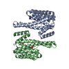
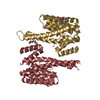
| Deposited unit | 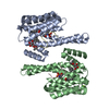
| ||||||||
|---|---|---|---|---|---|---|---|---|---|
| 1 |
| ||||||||
| Unit cell |
|
-Components
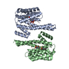
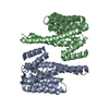
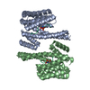
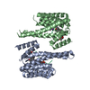
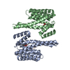
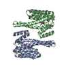
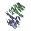
-Protein / Protein/peptide , 2 types, 4 molecules ABCD
| #1: Protein | Mass: 26316.764 Da / Num. of mol.: 2 Source method: isolated from a genetically manipulated source Source: (gene. exp.) Homo sapiens (human) / Gene: YWHAZ / Production host:  Escherichia coli (E. coli) / References: UniProt: P63104 Escherichia coli (E. coli) / References: UniProt: P63104#2: Protein/peptide | Mass: 1380.467 Da / Num. of mol.: 2 / Fragment: UNP residues 387-395 / Source method: obtained synthetically / Source: (synth.) Homo sapiens (human) / References: UniProt: Q9UQC2 |
|---|
-Non-polymers , 4 types, 198 molecules 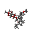


| #3: Chemical |  Mass: 536.697 Da / Num. of mol.: 2 / Source method: obtained synthetically / Formula: C30H48O8 Mass: 536.697 Da / Num. of mol.: 2 / Source method: obtained synthetically / Formula: C30H48O8#4: Chemical | Benzoic acid Mass: 122.121 Da / Num. of mol.: 2 / Source method: obtained synthetically / Formula: C7H6O2 Mass: 122.121 Da / Num. of mol.: 2 / Source method: obtained synthetically / Formula: C7H6O2#5: Chemical | ChemComp-MG / |  Mass: 24.305 Da / Num. of mol.: 1 / Source method: obtained synthetically / Formula: Mg / Source: (synth.) Homo sapiens (human) Mass: 24.305 Da / Num. of mol.: 1 / Source method: obtained synthetically / Formula: Mg / Source: (synth.) Homo sapiens (human)#6: Water | ChemComp-HOH / | WaterMass: 18.015 Da / Num. of mol.: 193 / Source method: isolated from a natural source / Formula: H2O |
|---|
-Experimental details
-Experiment
| Experiment | Method: X-RAY DIFFRACTION / Number of used crystals: 1 |
|---|
- Sample preparation
| Crystal | Density Matthews: 3.03 Å3/Da / Density % sol: 59.37 % |
|---|---|
| Crystal grow | Temperature: 277.15 K / Method: vapor diffusion, hanging drop Details: 0.17 M ammonium acetate, 0.085 M sodium citrate pH 5.6, 25.5% (w/v) PEG 4000, 15% (v/v) glycerol. |
-Data collection
| Diffraction | Mean temperature: 100 K | ||||||||||||||||||||||||||||||||||||||||||||||||||||||||||||||||||||||||||||||||||||||||||||||||||||||||||||||||||||||||||||||||||||||||||||||||||||||
|---|---|---|---|---|---|---|---|---|---|---|---|---|---|---|---|---|---|---|---|---|---|---|---|---|---|---|---|---|---|---|---|---|---|---|---|---|---|---|---|---|---|---|---|---|---|---|---|---|---|---|---|---|---|---|---|---|---|---|---|---|---|---|---|---|---|---|---|---|---|---|---|---|---|---|---|---|---|---|---|---|---|---|---|---|---|---|---|---|---|---|---|---|---|---|---|---|---|---|---|---|---|---|---|---|---|---|---|---|---|---|---|---|---|---|---|---|---|---|---|---|---|---|---|---|---|---|---|---|---|---|---|---|---|---|---|---|---|---|---|---|---|---|---|---|---|---|---|---|---|---|---|
| Diffraction source | Source: SYNCHROTRON / Site: SLS  / Beamline: X10SA / Wavelength: 1 Å / Beamline: X10SA / Wavelength: 1 Å | ||||||||||||||||||||||||||||||||||||||||||||||||||||||||||||||||||||||||||||||||||||||||||||||||||||||||||||||||||||||||||||||||||||||||||||||||||||||
| Detector | Type: DECTRIS PILATUS 6M / Detector: PIXEL / Date: Feb 10, 2012 | ||||||||||||||||||||||||||||||||||||||||||||||||||||||||||||||||||||||||||||||||||||||||||||||||||||||||||||||||||||||||||||||||||||||||||||||||||||||
| Radiation | Protocol: SINGLE WAVELENGTH / Monochromatic (M) / Laue (L): M / Scattering type: x-ray | ||||||||||||||||||||||||||||||||||||||||||||||||||||||||||||||||||||||||||||||||||||||||||||||||||||||||||||||||||||||||||||||||||||||||||||||||||||||
| Radiation wavelength | Wavelength: 1 Å / Relative weight: 1 | ||||||||||||||||||||||||||||||||||||||||||||||||||||||||||||||||||||||||||||||||||||||||||||||||||||||||||||||||||||||||||||||||||||||||||||||||||||||
| Reflection | Highest resolution: 1.95 Å / Num. obs: 49195 / % possible obs: 99.8 % / Observed criterion σ(I): -3 / Biso Wilson estimate: 36.714 Å2 / Rmerge F obs: 0.064 / Rmerge(I) obs: 0.045 / Rrim(I) all: 0.051 / Χ2: 0.995 / Net I/σ(I): 21.35 / Num. measured all: 247516 | ||||||||||||||||||||||||||||||||||||||||||||||||||||||||||||||||||||||||||||||||||||||||||||||||||||||||||||||||||||||||||||||||||||||||||||||||||||||
| Reflection shell | Diffraction-ID: 1 / Rejects: 0
|
-Phasing
| Phasing | Method: molecular replacement | |||||||||
|---|---|---|---|---|---|---|---|---|---|---|
| Phasing MR | Model details: Phaser MODE: MR_AUTO
|
- Processing
| Software |
| |||||||||||||||||||||||||||||||||||||||||||||||||||||||||||||||||
|---|---|---|---|---|---|---|---|---|---|---|---|---|---|---|---|---|---|---|---|---|---|---|---|---|---|---|---|---|---|---|---|---|---|---|---|---|---|---|---|---|---|---|---|---|---|---|---|---|---|---|---|---|---|---|---|---|---|---|---|---|---|---|---|---|---|---|
| Refinement | Method to determine structure: MOLECULAR REPLACEMENT / Resolution: 1.95→48.87 Å / Cor.coef. Fo:Fc: 0.951 / Cor.coef. Fo:Fc free: 0.938 / WRfactor Rfree: 0.2372 / WRfactor Rwork: 0.1987 / FOM work R set: 0.8568 / SU B: 3.085 / SU ML: 0.091 / SU R Cruickshank DPI: 0.1448 / SU Rfree: 0.1376 / Cross valid method: THROUGHOUT / σ(F): 0 / ESU R: 0.145 / ESU R Free: 0.138 / Stereochemistry target values: MAXIMUM LIKELIHOOD Details: HYDROGENS HAVE BEEN ADDED IN THE RIDING POSITIONS U VALUES : REFINED INDIVIDUALLY
| |||||||||||||||||||||||||||||||||||||||||||||||||||||||||||||||||
| Solvent computation | Ion probe radii: 0.8 Å / Shrinkage radii: 0.8 Å / VDW probe radii: 1.4 Å / Solvent model: MASK | |||||||||||||||||||||||||||||||||||||||||||||||||||||||||||||||||
| Displacement parameters | Biso max: 269.21 Å2 / Biso mean: 31.043 Å2 / Biso min: 13.87 Å2
| |||||||||||||||||||||||||||||||||||||||||||||||||||||||||||||||||
| Refinement step | Cycle: final / Resolution: 1.95→48.87 Å
| |||||||||||||||||||||||||||||||||||||||||||||||||||||||||||||||||
| Refine LS restraints |
| |||||||||||||||||||||||||||||||||||||||||||||||||||||||||||||||||
| LS refinement shell | Resolution: 1.95→2.001 Å / Total num. of bins used: 20
|
