 Movie
Movie Controller

[English] 日本語
 Yorodumi
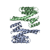
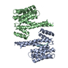
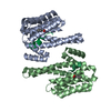
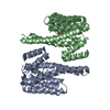
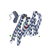
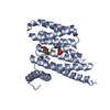
- PDB-4fj3: 14-3-3 isoform zeta in complex with a diphoyphorylated C-RAF peptide -
+ Open data

- Basic information
| Entry | Database: PDB / ID: 4fj3 | ||||||
|---|---|---|---|---|---|---|---|
| Title | 14-3-3 isoform zeta in complex with a diphoyphorylated C-RAF peptide | ||||||
 Components Components |
| ||||||
 Keywords Keywords | PROTEIN BINDING/TRANSFERASE / 14-3-3 fold / all alpha-helical /  adapter protein / PROTEIN BINDING-TRANSFERASE complex adapter protein / PROTEIN BINDING-TRANSFERASE complex | ||||||
| Function / homology |  Function and homology informationdeath-inducing signaling complex assembly / Golgi reassembly / intermediate filament cytoskeleton organization / regulation of synapse maturation / NOTCH4 Activation and Transmission of Signal to the Nucleus / establishment of Golgi localization / type B pancreatic cell proliferation / regulation of Rho protein signal transduction / SHOC2 M1731 mutant abolishes MRAS complex function / Gain-of-function MRAS complexes activate RAF signaling ...death-inducing signaling complex assembly / Golgi reassembly / intermediate filament cytoskeleton organization / regulation of synapse maturation / NOTCH4 Activation and Transmission of Signal to the Nucleus / establishment of Golgi localization / type B pancreatic cell proliferation / regulation of Rho protein signal transduction / SHOC2 M1731 mutant abolishes MRAS complex function / Gain-of-function MRAS complexes activate RAF signaling / Rap1 signalling / regulation of cell motility / insulin secretion involved in cellular response to glucose stimulus / negative regulation of protein localization to nucleus / Negative feedback regulation of MAPK pathway / KSRP (KHSRP) binds and destabilizes mRNA / IFNG signaling activates MAPKs / GP1b-IX-V activation signalling / ERBB2-ERBB3 signaling pathway / regulation of cell differentiation / face development / pseudopodium / somatic stem cell population maintenance / neurotrophin TRK receptor signaling pathway / thyroid gland development / Regulation of localization of FOXO transcription factors / Interleukin-3, Interleukin-5 and GM-CSF signaling / extrinsic apoptotic signaling pathway via death domain receptors / MAP kinase kinase kinase activity / phosphoserine residue binding / Activation of BAD and translocation to mitochondria / negative regulation of protein-containing complex assembly / SARS-CoV-2 targets host intracellular signalling and regulatory pathways / cellular response to glucose starvation / Chk1/Chk2(Cds1) mediated inactivation of Cyclin B:Cdk1 complex / Schwann cell development / type II interferon-mediated signaling pathway / SARS-CoV-1 targets host intracellular signalling and regulatory pathways / negative regulation of extrinsic apoptotic signaling pathway via death domain receptors / RHO GTPases activate PKNs / negative regulation of TORC1 signaling / activation of adenylate cyclase activity / response to muscle stretch / myelination / negative regulation of innate immune response / CD209 (DC-SIGN) signaling / regulation of ERK1 and ERK2 cascade / protein sequestering activity / insulin-like growth factor receptor signaling pathway / thymus development / Translocation of SLC2A4 (GLUT4) to the plasma membrane / Deactivation of the beta-catenin transactivating complex / TP53 Regulates Metabolic Genes / Negative regulation of NOTCH4 signaling / RAF activation / Signaling by high-kinase activity BRAF mutants / MAP2K and MAPK activation / wound healing / negative regulation of cysteine-type endopeptidase activity involved in apoptotic process / Stimuli-sensing channels / Negative regulation of MAPK pathway / Signaling by RAF1 mutants / Signaling by moderate kinase activity BRAF mutants / Paradoxical activation of RAF signaling by kinase inactive BRAF / Signaling downstream of RAS mutants / MAPK cascade / Signaling by BRAF and RAF1 fusions / melanosome / insulin receptor signaling pathway / positive regulation of peptidyl-serine phosphorylation / regulation of apoptotic process / DNA-binding transcription factor binding / blood microparticle / vesicle / mitochondrial outer membrane / transmembrane transporter binding / positive regulation of MAPK cascade / non-specific serine/threonine protein kinase / protein kinase activity / cadherin binding / negative regulation of cell population proliferation / protein phosphorylation / focal adhesion / protein serine kinase activity / protein serine/threonine kinase activity / glutamatergic synapse / apoptotic process / ubiquitin protein ligase binding / negative regulation of apoptotic process / protein kinase binding / Golgi apparatus / negative regulation of transcription by RNA polymerase II / enzyme binding / signal transduction / positive regulation of transcription by RNA polymerase II / mitochondrion / extracellular space / RNA binding / extracellular exosome / nucleoplasm Function and homology informationdeath-inducing signaling complex assembly / Golgi reassembly / intermediate filament cytoskeleton organization / regulation of synapse maturation / NOTCH4 Activation and Transmission of Signal to the Nucleus / establishment of Golgi localization / type B pancreatic cell proliferation / regulation of Rho protein signal transduction / SHOC2 M1731 mutant abolishes MRAS complex function / Gain-of-function MRAS complexes activate RAF signaling ...death-inducing signaling complex assembly / Golgi reassembly / intermediate filament cytoskeleton organization / regulation of synapse maturation / NOTCH4 Activation and Transmission of Signal to the Nucleus / establishment of Golgi localization / type B pancreatic cell proliferation / regulation of Rho protein signal transduction / SHOC2 M1731 mutant abolishes MRAS complex function / Gain-of-function MRAS complexes activate RAF signaling / Rap1 signalling / regulation of cell motility / insulin secretion involved in cellular response to glucose stimulus / negative regulation of protein localization to nucleus / Negative feedback regulation of MAPK pathway / KSRP (KHSRP) binds and destabilizes mRNA / IFNG signaling activates MAPKs / GP1b-IX-V activation signalling / ERBB2-ERBB3 signaling pathway / regulation of cell differentiation / face development / pseudopodium / somatic stem cell population maintenance / neurotrophin TRK receptor signaling pathway / thyroid gland development / Regulation of localization of FOXO transcription factors / Interleukin-3, Interleukin-5 and GM-CSF signaling / extrinsic apoptotic signaling pathway via death domain receptors / MAP kinase kinase kinase activity / phosphoserine residue binding / Activation of BAD and translocation to mitochondria / negative regulation of protein-containing complex assembly / SARS-CoV-2 targets host intracellular signalling and regulatory pathways / cellular response to glucose starvation / Chk1/Chk2(Cds1) mediated inactivation of Cyclin B:Cdk1 complex / Schwann cell development / type II interferon-mediated signaling pathway / SARS-CoV-1 targets host intracellular signalling and regulatory pathways / negative regulation of extrinsic apoptotic signaling pathway via death domain receptors / RHO GTPases activate PKNs / negative regulation of TORC1 signaling / activation of adenylate cyclase activity / response to muscle stretch / myelination / negative regulation of innate immune response / CD209 (DC-SIGN) signaling / regulation of ERK1 and ERK2 cascade / protein sequestering activity / insulin-like growth factor receptor signaling pathway / thymus development / Translocation of SLC2A4 (GLUT4) to the plasma membrane / Deactivation of the beta-catenin transactivating complex / TP53 Regulates Metabolic Genes / Negative regulation of NOTCH4 signaling / RAF activation / Signaling by high-kinase activity BRAF mutants / MAP2K and MAPK activation / wound healing / negative regulation of cysteine-type endopeptidase activity involved in apoptotic process / Stimuli-sensing channels / Negative regulation of MAPK pathway / Signaling by RAF1 mutants / Signaling by moderate kinase activity BRAF mutants / Paradoxical activation of RAF signaling by kinase inactive BRAF / Signaling downstream of RAS mutants / MAPK cascade / Signaling by BRAF and RAF1 fusions / melanosome / insulin receptor signaling pathway / positive regulation of peptidyl-serine phosphorylation / regulation of apoptotic process / DNA-binding transcription factor binding / blood microparticle / vesicle / mitochondrial outer membrane / transmembrane transporter binding / positive regulation of MAPK cascade / non-specific serine/threonine protein kinase / protein kinase activity / cadherin binding / negative regulation of cell population proliferation / protein phosphorylation / focal adhesion / protein serine kinase activity / protein serine/threonine kinase activity / glutamatergic synapse / apoptotic process / ubiquitin protein ligase binding / negative regulation of apoptotic process / protein kinase binding / Golgi apparatus / negative regulation of transcription by RNA polymerase II / enzyme binding / signal transduction / positive regulation of transcription by RNA polymerase II / mitochondrion / extracellular space / RNA binding / extracellular exosome / nucleoplasmSimilarity search - Function | ||||||
| Biological species |  Homo sapiens (human) Homo sapiens (human) | ||||||
| Method | X-RAY DIFFRACTION / SYNCHROTRON / MOLECULAR REPLACEMENT / molecular replacement / Resolution: 1.95 Å | ||||||
 Authors Authors | Ottmann, C. / Molzan, M. | ||||||
 Citation Citation | Journal: J.Mol.Biol. / Year: 2012 Title: Synergistic binding of the phosphorylated S233- and S259-binding sites of C-RAF to one 14-3-3zeta dimer Authors: Molzan, M. / Ottmann, C. | ||||||
| History |
|
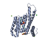
- Structure visualization
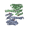
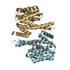
| Structure viewer | Molecule: MolmilJmol/JSmol |
|---|
- Downloads & links
-Download
| PDBx/mmCIF format | 4fj3.cif.gz | 221.3 KB | Display | PDBx/mmCIF format |
|---|---|---|---|---|
| PDB format | pdb4fj3.ent.gz | 185.1 KB | Display | PDB format |
| PDBx/mmJSON format | 4fj3.json.gz | Tree view | PDBx/mmJSON format | |
| Others |  Other downloads Other downloads |
-Validation report
| Arichive directory | https://data.pdbj.org/pub/pdb/validation_reports/fj/4fj3ftp://data.pdbj.org/pub/pdb/validation_reports/fj/4fj3 | HTTPS FTP |
|---|
-Related structure data
| Related structure data | |
|---|---|
| Similar structure data |

-Links
 PDBj
PDBj


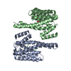
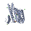
- Assembly
| Deposited unit | 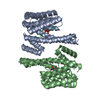
| |||||||||||||||||||||||||||
|---|---|---|---|---|---|---|---|---|---|---|---|---|---|---|---|---|---|---|---|---|---|---|---|---|---|---|---|---|
| 1 |
| |||||||||||||||||||||||||||
| Unit cell |
| |||||||||||||||||||||||||||
| Noncrystallographic symmetry (NCS) | NCS domain:
NCS domain segments:
|
-Components
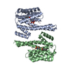
| #1: Protein | Mass: 26720.217 Da / Num. of mol.: 2 / Fragment: UNP residues 1-230 Source method: isolated from a genetically manipulated source Source: (gene. exp.) Homo sapiens (human) / Gene: YWHAZ / Production host:  Escherichia coli (E. coli) / References: UniProt: P63104 Escherichia coli (E. coli) / References: UniProt: P63104#2: Protein/peptide | | Mass: 4184.204 Da / Num. of mol.: 1 / Fragment: UNP residues 229-264 / Source method: obtained synthetically / Details: synthetic peptide / Source: (synth.) Homo sapiens (human)References: UniProt: P04049, non-specific serine/threonine protein kinase#3: Water | ChemComp-HOH / | Water Mass: 18.015 Da / Num. of mol.: 587 / Source method: isolated from a natural source / Formula: H2O Mass: 18.015 Da / Num. of mol.: 587 / Source method: isolated from a natural source / Formula: H2O |
|---|
-Experimental details
-Experiment
| Experiment | Method: X-RAY DIFFRACTION / Number of used crystals: 1 |
|---|
- Sample preparation
| Crystal | Density Matthews: 3.65 Å3/Da / Density % sol: 66.31 % |
|---|---|
| Crystal grow | Temperature: 277 K / Method: vapor diffusion, sitting drop / pH: 7 Details: 0.1 M Na-acetate pH 7.0, 0.8 M NaH2PO4 and 1.2 M K2HPO4 , VAPOR DIFFUSION, SITTING DROP, temperature 277K |
-Data collection
| Diffraction | Mean temperature: 100 K | |||||||||||||||||||||||||||||||||||||||||||||||||||||||||||||||||||||||||||||
|---|---|---|---|---|---|---|---|---|---|---|---|---|---|---|---|---|---|---|---|---|---|---|---|---|---|---|---|---|---|---|---|---|---|---|---|---|---|---|---|---|---|---|---|---|---|---|---|---|---|---|---|---|---|---|---|---|---|---|---|---|---|---|---|---|---|---|---|---|---|---|---|---|---|---|---|---|---|---|
| Diffraction source | Source: SYNCHROTRON / Site: SLS  / Beamline: X10SA / Wavelength: 0.9778 Å / Beamline: X10SA / Wavelength: 0.9778 Å | |||||||||||||||||||||||||||||||||||||||||||||||||||||||||||||||||||||||||||||
| Detector | Type: PSI PILATUS 6M / Detector: PIXEL / Date: Feb 14, 2011 | |||||||||||||||||||||||||||||||||||||||||||||||||||||||||||||||||||||||||||||
| Radiation | Monochromator: Al2/Al2 / Protocol: SINGLE WAVELENGTH / Monochromatic (M) / Laue (L): M / Scattering type: x-ray | |||||||||||||||||||||||||||||||||||||||||||||||||||||||||||||||||||||||||||||
| Radiation wavelength | Wavelength: 0.9778 Å / Relative weight: 1 | |||||||||||||||||||||||||||||||||||||||||||||||||||||||||||||||||||||||||||||
| Reflection | Resolution: 1.95→50 Å / Num. all: 62201 / Num. obs: 61869 / % possible obs: 99.5 % / Observed criterion σ(F): -3 / Observed criterion σ(I): -3 / Biso Wilson estimate: 36.724 Å2 / Rmerge(I) obs: 0.052 / Net I/σ(I): 19.57 | |||||||||||||||||||||||||||||||||||||||||||||||||||||||||||||||||||||||||||||
| Reflection shell |
|
-Phasing
| Phasing | Method: molecular replacement |
|---|
- Processing
| Software |
| ||||||||||||||||||||||||||||||||||||||||||||||||||||||||||||
|---|---|---|---|---|---|---|---|---|---|---|---|---|---|---|---|---|---|---|---|---|---|---|---|---|---|---|---|---|---|---|---|---|---|---|---|---|---|---|---|---|---|---|---|---|---|---|---|---|---|---|---|---|---|---|---|---|---|---|---|---|---|
| Refinement | Method to determine structure: MOLECULAR REPLACEMENT / Resolution: 1.95→46.73 Å / Cor.coef. Fo:Fc: 0.962 / Cor.coef. Fo:Fc free: 0.947 / Occupancy max: 1 / Occupancy min: 0.2 / SU B: 5.167 / SU ML: 0.068 / Cross valid method: THROUGHOUT / σ(F): 4 / ESU R: 0.15 / ESU R Free: 0.113 / Stereochemistry target values: MAXIMUM LIKELIHOOD Details: HYDROGENS HAVE BEEN USED IF PRESENT IN THE INPUT U VALUES : REFINED INDIVIDUALLY
| ||||||||||||||||||||||||||||||||||||||||||||||||||||||||||||
| Solvent computation | Ion probe radii: 0.8 Å / Shrinkage radii: 0.8 Å / VDW probe radii: 1.2 Å / Solvent model: MASK | ||||||||||||||||||||||||||||||||||||||||||||||||||||||||||||
| Displacement parameters | Biso max: 87.56 Å2 / Biso mean: 35.2967 Å2 / Biso min: 14.65 Å2
| ||||||||||||||||||||||||||||||||||||||||||||||||||||||||||||
| Refinement step | Cycle: LAST / Resolution: 1.95→46.73 Å
| ||||||||||||||||||||||||||||||||||||||||||||||||||||||||||||
| Refine LS restraints |
| ||||||||||||||||||||||||||||||||||||||||||||||||||||||||||||
| Refine LS restraints NCS | Dom-ID: 1 / Auth asym-ID: A / Ens-ID: 1 / Refine-ID: X-RAY DIFFRACTION
| ||||||||||||||||||||||||||||||||||||||||||||||||||||||||||||
| LS refinement shell | Resolution: 1.95→2.001 Å / Total num. of bins used: 20
|
