 Movie
Movie Controller


+ Open data

- Basic information
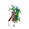
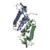
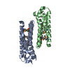
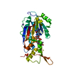
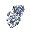
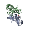
| Entry | Database: PDB / ID: 5en1 | ||||||
|---|---|---|---|---|---|---|---|
| Title | Crystal structure of hnRNPA2B1 in complex with RNA | ||||||
 Components Components |
| ||||||
 Keywords Keywords | RNA BINDING PROTEIN/RNA /  HNRNP / RRM / RNA BINDING PROTEIN-RNA complex HNRNP / RRM / RNA BINDING PROTEIN-RNA complex | ||||||
| Function / homology |  Function and homology information Function and homology informationpositive regulation of telomerase RNA reverse transcriptase activity / miRNA transport / positive regulation of telomere maintenance via telomere lengthening / RNA transport / G-quadruplex DNA unwinding / primary miRNA processing / single-stranded telomeric DNA binding / N6-methyladenosine-containing RNA reader activity / G-rich strand telomeric DNA binding / miRNA binding ...positive regulation of telomerase RNA reverse transcriptase activity / miRNA transport / positive regulation of telomere maintenance via telomere lengthening / RNA transport / G-quadruplex DNA unwinding / primary miRNA processing / single-stranded telomeric DNA binding / N6-methyladenosine-containing RNA reader activity / G-rich strand telomeric DNA binding / miRNA binding / negative regulation of mRNA splicing, via spliceosome / Processing of Capped Intron-Containing Pre-mRNA / mRNA transport / Cajal body / mRNA export from nucleus / pre-mRNA intronic binding / Gene and protein expression by JAK-STAT signaling after Interleukin-12 stimulation / catalytic step 2 spliceosome / molecular condensate scaffold activity / mRNA Splicing - Major Pathway / mRNA 3'-UTR binding / spliceosomal complex / mRNA processing / mRNA splicing, via spliceosome / nuclear matrix / chromosome, telomeric region / ribonucleoprotein complex / negative regulation of transcription by RNA polymerase II / RNA binding / extracellular exosome / nucleoplasm / membrane / identical protein binding / nucleus / cytoplasmSimilarity search - Function | ||||||
| Biological species |  Homo sapiens (human) Homo sapiens (human) | ||||||
| Method | X-RAY DIFFRACTION / SYNCHROTRON / MOLECULAR REPLACEMENT / Resolution: 2.58 Å | ||||||
 Authors Authors | Wu, B.X. / Su, S.C. / Ma, J.B. | ||||||
 Citation Citation | Journal: Nat Commun / Year: 2018 Title: Molecular basis for the specific and multivariant recognitions of RNA substrates by human hnRNP A2/B1 Authors: Wu, B.X. / Su, S.C. / Patil, D.P. / Liu, H.H. / Gan, J.H. / Jaffrey, S.R. / Ma, J.B. | ||||||
| History |
|
- Structure visualization
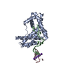
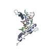
| Structure viewer | Molecule: MolmilJmol/JSmol |
|---|
- Downloads & links
-Download
| PDBx/mmCIF format | 5en1.cif.gz | 56.7 KB | Display | PDBx/mmCIF format |
|---|---|---|---|---|
| PDB format | pdb5en1.ent.gz | 38.1 KB | Display | PDB format |
| PDBx/mmJSON format | 5en1.json.gz | Tree view | PDBx/mmJSON format | |
| Others |  Other downloads Other downloads |
-Validation report
| Arichive directory | https://data.pdbj.org/pub/pdb/validation_reports/en/5en1ftp://data.pdbj.org/pub/pdb/validation_reports/en/5en1 | HTTPS FTP |
|---|
-Related structure data
| Related structure data | 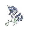 1u1qS S: Starting model for refinement |
|---|---|
| Similar structure data |

-Links
 PDBj
PDBj


- Assembly
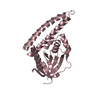
| Deposited unit | 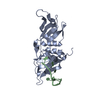
| ||||||||
|---|---|---|---|---|---|---|---|---|---|
| 1 |
| ||||||||
| Unit cell |
|
-Components
| #1: Protein | Mass: 21206.969 Da / Num. of mol.: 1 / Fragment: UNP residues 12-195 Source method: isolated from a genetically manipulated source Source: (gene. exp.) Homo sapiens (human) / Gene: HNRNPA2B1, HNRPA2B1 / Production host:  Escherichia coli (E. coli) / References: UniProt: P22626 Escherichia coli (E. coli) / References: UniProt: P22626 |
|---|---|
| #2: RNA chain | Mass: 2260.419 Da / Num. of mol.: 1 / Source method: obtained synthetically / Source: (synth.) Homo sapiens (human) |
| #3: Water | ChemComp-HOH / Water Mass: 18.015 Da / Num. of mol.: 11 / Source method: isolated from a natural source / Formula: H2O Mass: 18.015 Da / Num. of mol.: 11 / Source method: isolated from a natural source / Formula: H2O |
-Experimental details
-Experiment
| Experiment | Method: X-RAY DIFFRACTION |
|---|
- Sample preparation
| Crystal | Density Matthews: 2.26 Å3/Da / Density % sol: 45.59 % |
|---|---|
| Crystal grow | Temperature: 293 K / Method: vapor diffusion, sitting drop / pH: 7 / Details: 25% PEG3350, 0.1M Tris pH8.5 |
-Data collection
| Diffraction | Mean temperature: 100 K |
|---|---|
| Diffraction source | Source: SYNCHROTRON / Site: SSRF  / Beamline: BL18U1 / Wavelength: 0.987 Å / Beamline: BL18U1 / Wavelength: 0.987 Å |
| Detector | Type: RIGAKU / Detector: CCD / Date: Oct 26, 2015 |
| Radiation | Protocol: SINGLE WAVELENGTH / Monochromatic (M) / Laue (L): M / Scattering type: x-ray |
| Radiation wavelength | Wavelength: 0.987 Å / Relative weight: 1 |
| Reflection | Resolution: 2.58→28.81 Å / Num. obs: 6187 / % possible obs: 91.89 % / Redundancy: 2.5 % / Net I/σ(I): 7.9 |
- Processing
| Software |
| ||||||||||||||||||||||||||||||||||||||||||||||||||||||||||||||||||||||||||||||||||||||||||||||||||||||||||||||||||||||||||||||||||||||||||||||||||||||||||||||||||||||||||||||||||||||
|---|---|---|---|---|---|---|---|---|---|---|---|---|---|---|---|---|---|---|---|---|---|---|---|---|---|---|---|---|---|---|---|---|---|---|---|---|---|---|---|---|---|---|---|---|---|---|---|---|---|---|---|---|---|---|---|---|---|---|---|---|---|---|---|---|---|---|---|---|---|---|---|---|---|---|---|---|---|---|---|---|---|---|---|---|---|---|---|---|---|---|---|---|---|---|---|---|---|---|---|---|---|---|---|---|---|---|---|---|---|---|---|---|---|---|---|---|---|---|---|---|---|---|---|---|---|---|---|---|---|---|---|---|---|---|---|---|---|---|---|---|---|---|---|---|---|---|---|---|---|---|---|---|---|---|---|---|---|---|---|---|---|---|---|---|---|---|---|---|---|---|---|---|---|---|---|---|---|---|---|---|---|---|---|
| Refinement | Method to determine structure: MOLECULAR REPLACEMENT Starting model: 1U1Q Resolution: 2.58→28.81 Å / Cor.coef. Fo:Fc: 0.94 / Cor.coef. Fo:Fc free: 0.925 / SU B: 14.648 / SU ML: 0.298 / Cross valid method: THROUGHOUT / ESU R Free: 0.338 / Stereochemistry target values: MAXIMUM LIKELIHOOD / Details: HYDROGENS HAVE BEEN ADDED IN THE RIDING POSITIONS
| ||||||||||||||||||||||||||||||||||||||||||||||||||||||||||||||||||||||||||||||||||||||||||||||||||||||||||||||||||||||||||||||||||||||||||||||||||||||||||||||||||||||||||||||||||||||
| Solvent computation | Ion probe radii: 0.8 Å / Shrinkage radii: 0.8 Å / VDW probe radii: 1.2 Å / Solvent model: MASK | ||||||||||||||||||||||||||||||||||||||||||||||||||||||||||||||||||||||||||||||||||||||||||||||||||||||||||||||||||||||||||||||||||||||||||||||||||||||||||||||||||||||||||||||||||||||
| Displacement parameters | Biso mean: 45.201 Å2
| ||||||||||||||||||||||||||||||||||||||||||||||||||||||||||||||||||||||||||||||||||||||||||||||||||||||||||||||||||||||||||||||||||||||||||||||||||||||||||||||||||||||||||||||||||||||
| Refinement step | Cycle: 1 / Resolution: 2.58→28.81 Å
| ||||||||||||||||||||||||||||||||||||||||||||||||||||||||||||||||||||||||||||||||||||||||||||||||||||||||||||||||||||||||||||||||||||||||||||||||||||||||||||||||||||||||||||||||||||||
| Refine LS restraints |
|