 Movie
Movie Controller


+ Open data

- Basic information
| Entry | Database: PDB / ID: 3a8p | ||||||
|---|---|---|---|---|---|---|---|
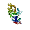
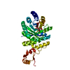
| Title | Crystal structure of the Tiam2 PHCCEx domain | ||||||
 Components Components | T-lymphoma invasion and metastasis-inducing protein 2 | ||||||
 Keywords Keywords |  SIGNALING PROTEIN / guanine nucleotide exchange factor / Alternative splicing / Cell projection / Coiled coil / Cytoplasm / Guanine-nucleotide releasing factor / Lipoprotein / Myristate / Phosphoprotein SIGNALING PROTEIN / guanine nucleotide exchange factor / Alternative splicing / Cell projection / Coiled coil / Cytoplasm / Guanine-nucleotide releasing factor / Lipoprotein / Myristate / Phosphoprotein | ||||||
| Function / homology |  Function and homology information Function and homology informationNRAGE signals death through JNK / RAC1 GTPase cycle / G alpha (12/13) signalling events / positive regulation of axonogenesis / small GTPase-mediated signal transduction / GTPase activator activity / guanyl-nucleotide exchange factor activity / filopodium / lamellipodium / growth cone ...NRAGE signals death through JNK / RAC1 GTPase cycle / G alpha (12/13) signalling events / positive regulation of axonogenesis / small GTPase-mediated signal transduction / GTPase activator activity / guanyl-nucleotide exchange factor activity / filopodium / lamellipodium / growth cone / perikaryon / synapse / membrane / cytoplasmSimilarity search - Function | ||||||
| Biological species |  Mus musculus (house mouse) Mus musculus (house mouse) | ||||||
| Method | X-RAY DIFFRACTION / SYNCHROTRON / MOLECULAR REPLACEMENT / Resolution: 2.1 Å | ||||||
 Authors Authors | Terawaki, S. / Kitano, K. / Mori, T. / Zhai, Y. / Higuchi, Y. / Itoh, N. / Watanabe, T. / Kaibuchi, K. / Hakoshima, T. | ||||||
 Citation Citation | Journal: Embo J. / Year: 2010 Title: The PHCCEx domain of Tiam1/2 is a novel protein- and membrane-binding module Authors: Terawaki, S. / Kitano, K. / Mori, T. / Zhai, Y. / Higuchi, Y. / Itoh, N. / Watanabe, T. / Kaibuchi, K. / Hakoshima, T. | ||||||
| History |
|
- Structure visualization
| Structure viewer | Molecule: MolmilJmol/JSmol |
|---|
- Downloads & links
-Download
| PDBx/mmCIF format | 3a8p.cif.gz | 198.5 KB | Display | PDBx/mmCIF format |
|---|---|---|---|---|
| PDB format | pdb3a8p.ent.gz | 158.1 KB | Display | PDB format |
| PDBx/mmJSON format | 3a8p.json.gz | Tree view | PDBx/mmJSON format | |
| Others |  Other downloads Other downloads |
-Validation report
| Arichive directory | https://data.pdbj.org/pub/pdb/validation_reports/a8/3a8pftp://data.pdbj.org/pub/pdb/validation_reports/a8/3a8p | HTTPS FTP |
|---|
-Related structure data
| Related structure data | 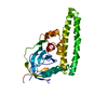 3a8nC  3a8qSC C: citing same article ( S: Starting model for refinement |
|---|---|
| Similar structure data |


-Links
 PDBj
PDBj


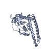
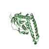
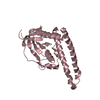
- Assembly
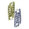
| Deposited unit | 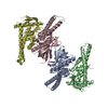
| ||||||||
|---|---|---|---|---|---|---|---|---|---|
| 1 | 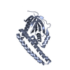
| ||||||||
| 2 | 
| ||||||||
| 3 | 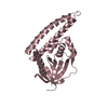
| ||||||||
| 4 | 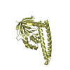
| ||||||||
| Unit cell |
|
-Components
| #1: Protein | Mass: 29925.717 Da / Num. of mol.: 4 / Fragment: PHCCEx domain, residues 500-757 Source method: isolated from a genetically manipulated source Source: (gene. exp.) Mus musculus (house mouse) / Gene: Tiam2, Kiaa2016, Stef / Plasmid: pGEX6P-3 / Production host:  Escherichia coli (E. coli) / Strain (production host): BL21RIL / References: UniProt: Q6ZPF3 Escherichia coli (E. coli) / Strain (production host): BL21RIL / References: UniProt: Q6ZPF3#2: Water | ChemComp-HOH / | Water Mass: 18.015 Da / Num. of mol.: 383 / Source method: isolated from a natural source / Formula: H2O Mass: 18.015 Da / Num. of mol.: 383 / Source method: isolated from a natural source / Formula: H2O |
|---|
-Experimental details
-Experiment
| Experiment | Method: X-RAY DIFFRACTION / Number of used crystals: 1 |
|---|
- Sample preparation
| Crystal | Density Matthews: 2.34 Å3/Da / Density % sol: 47.39 % |
|---|---|
| Crystal grow | Temperature: 293 K / Method: vapor diffusion, hanging drop / pH: 8.5 Details: 20% PEG 8000, 100mM Tris-HCl, 200mM Magnesium chloride, pH 8.5, VAPOR DIFFUSION, HANGING DROP, temperature 293K |
-Data collection
| Diffraction | Mean temperature: 100 K |
|---|---|
| Diffraction source | Source: SYNCHROTRON / Site: SPring-8  / Beamline: BL41XU / Wavelength: 1 Å / Beamline: BL41XU / Wavelength: 1 Å |
| Detector | Type: ADSC QUANTUM 315 / Detector: CCD / Date: May 25, 2007 |
| Radiation | Monochromator: rotated-inclined double-crystal / Protocol: SINGLE WAVELENGTH / Monochromatic (M) / Laue (L): M / Scattering type: x-ray |
| Radiation wavelength | Wavelength: 1 Å / Relative weight: 1 |
| Reflection | Resolution: 2.08→50 Å / Num. obs: 64571 / % possible obs: 97.6 % / Redundancy: 3.6 % / Biso Wilson estimate: 22.8 Å2 |
| Reflection shell | Resolution: 2.08→2.15 Å / Redundancy: 2.9 % / Mean I/σ(I) obs: 1.9 / % possible all: 80.6 |
- Processing
| Software |
| ||||||||||||||||||||||||||||||||||||||||||||||||||||||||||||
|---|---|---|---|---|---|---|---|---|---|---|---|---|---|---|---|---|---|---|---|---|---|---|---|---|---|---|---|---|---|---|---|---|---|---|---|---|---|---|---|---|---|---|---|---|---|---|---|---|---|---|---|---|---|---|---|---|---|---|---|---|---|
| Refinement | Method to determine structure: MOLECULAR REPLACEMENT Starting model: PDB ENTRY 3A8Q Resolution: 2.1→29.81 Å / Rfactor Rfree error: 0.003 / Data cutoff high absF: 1845113.44 / Data cutoff low absF: 0 / Isotropic thermal model: RESTRAINED / Cross valid method: THROUGHOUT / σ(F): 0 / Details: BULK SOLVENT MODEL USED
| ||||||||||||||||||||||||||||||||||||||||||||||||||||||||||||
| Solvent computation | Solvent model: FLAT MODEL / Bsol: 60.7651 Å2 / ksol: 0.4 e/Å3 | ||||||||||||||||||||||||||||||||||||||||||||||||||||||||||||
| Displacement parameters | Biso mean: 39.2 Å2
| ||||||||||||||||||||||||||||||||||||||||||||||||||||||||||||
| Refine analyze |
| ||||||||||||||||||||||||||||||||||||||||||||||||||||||||||||
| Refinement step | Cycle: LAST / Resolution: 2.1→29.81 Å
| ||||||||||||||||||||||||||||||||||||||||||||||||||||||||||||
| Refine LS restraints |
| ||||||||||||||||||||||||||||||||||||||||||||||||||||||||||||
| LS refinement shell | Resolution: 2.1→2.23 Å / Rfactor Rfree error: 0.01 / Total num. of bins used: 6
| ||||||||||||||||||||||||||||||||||||||||||||||||||||||||||||
| Xplor file |
|
