 Movie
Movie Controller


+ Open data

- Basic information
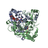
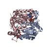
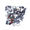
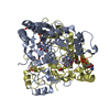
| Entry | Database: PDB / ID: 5ea2 | ||||||
|---|---|---|---|---|---|---|---|
| Title | Crystal Structure of Holo NAD(P)H dehydrogenase, quinone 1 | ||||||
 Components Components | NAD(P)H dehydrogenase [quinone] 1 | ||||||
 Keywords Keywords |  OXIDOREDUCTASE / NQO1 / two-electron reduction of Quinone / NAD(P)H dehydrogenase OXIDOREDUCTASE / NQO1 / two-electron reduction of Quinone / NAD(P)H dehydrogenase | ||||||
| Function / homology |  Function and homology information Function and homology informationresponse to L-glutamine / response to flavonoid / ubiquinone metabolic process / vitamin E metabolic process / vitamin K metabolic process / NADPH dehydrogenase (quinone) activity / NAD(P)H dehydrogenase (quinone) / NADH oxidation / cellular response to metal ion / cytochrome-b5 reductase activity, acting on NAD(P)H ...response to L-glutamine / response to flavonoid / ubiquinone metabolic process / vitamin E metabolic process / vitamin K metabolic process / NADPH dehydrogenase (quinone) activity / NAD(P)H dehydrogenase (quinone) / NADH oxidation / cellular response to metal ion / cytochrome-b5 reductase activity, acting on NAD(P)H / NADPH oxidation / response to tetrachloromethane / NADH:ubiquinone reductase (non-electrogenic) activity / NAD(P)H dehydrogenase (quinone) activity / response to hydrogen sulfide / response to alkaloid / response to carbohydrate / synaptic transmission, cholinergic / Regulation of ornithine decarboxylase (ODC) / NFE2L2 regulating anti-oxidant/detoxification enzymes / response to testosterone / response to amine / superoxide dismutase activity / response to electrical stimulus / nitric oxide biosynthetic process / xenobiotic metabolic process / removal of superoxide radicals / response to nutrient / cell redox homeostasis / response to hormone / response to ischemia / protein catabolic process / negative regulation of protein catabolic process / response to toxic substance / cellular response to hydrogen peroxide / protein polyubiquitination / positive regulation of neuron apoptotic process / response to estradiol / cellular response to oxidative stress / response to ethanol / response to oxidative stress / response to lipopolysaccharide / innate immune response / neuronal cell body / synapse / dendrite / negative regulation of apoptotic process / RNA binding / identical protein binding / nucleus / cytosol / cytoplasmSimilarity search - Function | ||||||
| Biological species |  Homo sapiens (human) Homo sapiens (human) | ||||||
| Method | X-RAY DIFFRACTION / MOLECULAR REPLACEMENT / Resolution: 2.01 Å | ||||||
 Authors Authors | Pidugu, L.S. / Mbimba, J.E. / Ahmad, M. / Pozharski, E. / Sausville, E.A. / Emadi, A. / Toth, E.A. | ||||||
 Citation Citation | Journal: Bmc Struct.Biol. / Year: 2016 Title: A direct interaction between NQO1 and a chemotherapeutic dimeric naphthoquinone. Authors: Pidugu, L.S. / Mbimba, J.C. / Ahmad, M. / Pozharski, E. / Sausville, E.A. / Emadi, A. / Toth, E.A. | ||||||
| History |
|
- Structure visualization
| Structure viewer | Molecule: MolmilJmol/JSmol |
|---|
- Downloads & links
-Download
| PDBx/mmCIF format | 5ea2.cif.gz | 246.5 KB | Display | PDBx/mmCIF format |
|---|---|---|---|---|
| PDB format | pdb5ea2.ent.gz | 195.7 KB | Display | PDB format |
| PDBx/mmJSON format | 5ea2.json.gz | Tree view | PDBx/mmJSON format | |
| Others |  Other downloads Other downloads |
-Validation report
| Arichive directory | https://data.pdbj.org/pub/pdb/validation_reports/ea/5ea2ftp://data.pdbj.org/pub/pdb/validation_reports/ea/5ea2 | HTTPS FTP |
|---|
-Related structure data
| Related structure data |  5eaiC  1d4aS C: citing same article ( S: Starting model for refinement |
|---|---|
| Similar structure data |


-Links
 PDBj
PDBj
- Assembly
| Deposited unit | 
| ||||||||
|---|---|---|---|---|---|---|---|---|---|
| 1 | 
| ||||||||
| 2 | 
| ||||||||
| Unit cell |
|
-Components
| #1: Protein | Mass: 31070.742 Da / Num. of mol.: 4 Source method: isolated from a genetically manipulated source Source: (gene. exp.) Homo sapiens (human) / Gene: NQO1, DIA4, NMOR1 / Plasmid: 19-PPSDetails (production host): pET19b with a prescission protease cleave site inserted Production host:  Escherichia coli (E. coli) / Strain (production host): BL21 (DE3) Escherichia coli (E. coli) / Strain (production host): BL21 (DE3)References: UniProt: P15559, NAD(P)H dehydrogenase (quinone)#2: Chemical | ChemComp-FAD / Flavin adenine dinucleotide  Mass: 785.550 Da / Num. of mol.: 4 / Source method: obtained synthetically / Formula: C27H33N9O15P2 / Comment: FAD*YM Mass: 785.550 Da / Num. of mol.: 4 / Source method: obtained synthetically / Formula: C27H33N9O15P2 / Comment: FAD*YM#3: Water | ChemComp-HOH / | Water Mass: 18.015 Da / Num. of mol.: 713 / Source method: isolated from a natural source / Formula: H2O Mass: 18.015 Da / Num. of mol.: 713 / Source method: isolated from a natural source / Formula: H2O |
|---|
-Experimental details
-Experiment
| Experiment | Method: X-RAY DIFFRACTION / Number of used crystals: 1 |
|---|
- Sample preparation
| Crystal | Density Matthews: 2.42 Å3/Da / Density % sol: 49.14 % |
|---|---|
| Crystal grow | Temperature: 293 K / Method: vapor diffusion, sitting drop / pH: 7 / Details: PEG3350, Ammonium Citrate |
-Data collection
| Diffraction | Mean temperature: 100 K | |||||||||||||||||||||||||||
|---|---|---|---|---|---|---|---|---|---|---|---|---|---|---|---|---|---|---|---|---|---|---|---|---|---|---|---|---|
| Diffraction source | Source: ROTATING ANODE / Type: RIGAKU MICROMAX-007 / Wavelength: 1.54 Å | |||||||||||||||||||||||||||
| Detector | Type: RIGAKU RAXIS IV / Detector: IMAGE PLATE / Date: Feb 25, 2014 | |||||||||||||||||||||||||||
| Radiation | Protocol: SINGLE WAVELENGTH / Monochromatic (M) / Laue (L): M / Scattering type: x-ray | |||||||||||||||||||||||||||
| Radiation wavelength | Wavelength: 1.54 Å / Relative weight: 1 | |||||||||||||||||||||||||||
| Reflection | Resolution: 2.01→45.12 Å / Num. obs: 78225 / % possible obs: 100 % / Redundancy: 7.4 % / Biso Wilson estimate: 30.05 Å2 / Net I/σ(I): 7 | |||||||||||||||||||||||||||
| Reflection shell | Diffraction-ID: 1 / Rejects: 0
|
- Processing
| Software |
| ||||||||||||||||||||||||||||||||||||||||||||||||||||||||||||||||||||||||||||||||||||||||||||||||||||||||||||||||||
|---|---|---|---|---|---|---|---|---|---|---|---|---|---|---|---|---|---|---|---|---|---|---|---|---|---|---|---|---|---|---|---|---|---|---|---|---|---|---|---|---|---|---|---|---|---|---|---|---|---|---|---|---|---|---|---|---|---|---|---|---|---|---|---|---|---|---|---|---|---|---|---|---|---|---|---|---|---|---|---|---|---|---|---|---|---|---|---|---|---|---|---|---|---|---|---|---|---|---|---|---|---|---|---|---|---|---|---|---|---|---|---|---|---|---|---|
| Refinement | Method to determine structure: MOLECULAR REPLACEMENT Starting model: 1D4A Resolution: 2.01→44.57 Å / Cor.coef. Fo:Fc: 0.9441 / Cor.coef. Fo:Fc free: 0.9308 / SU R Cruickshank DPI: 0.178 / Cross valid method: THROUGHOUT / σ(F): 0 / SU R Blow DPI: 0.18 / SU Rfree Blow DPI: 0.152 / SU Rfree Cruickshank DPI: 0.152
| ||||||||||||||||||||||||||||||||||||||||||||||||||||||||||||||||||||||||||||||||||||||||||||||||||||||||||||||||||
| Displacement parameters | Biso mean: 28.94 Å2
| ||||||||||||||||||||||||||||||||||||||||||||||||||||||||||||||||||||||||||||||||||||||||||||||||||||||||||||||||||
| Refine analyze | Luzzati coordinate error obs: 0.223 Å | ||||||||||||||||||||||||||||||||||||||||||||||||||||||||||||||||||||||||||||||||||||||||||||||||||||||||||||||||||
| Refinement step | Cycle: LAST / Resolution: 2.01→44.57 Å
| ||||||||||||||||||||||||||||||||||||||||||||||||||||||||||||||||||||||||||||||||||||||||||||||||||||||||||||||||||
| Refine LS restraints |
| ||||||||||||||||||||||||||||||||||||||||||||||||||||||||||||||||||||||||||||||||||||||||||||||||||||||||||||||||||
| LS refinement shell | Resolution: 2.01→2.06 Å / Total num. of bins used: 20
|
