 Movie
Movie Controller

[English] 日本語
 Yorodumi
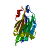
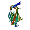
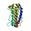
- PDB-5e8f: Structure of Fully modified geranylgeranylated PDE6C Peptide in c... -
+ Open data

- Basic information
| Entry | Database: PDB / ID: 5e8f | ||||||
|---|---|---|---|---|---|---|---|
| Title | Structure of Fully modified geranylgeranylated PDE6C Peptide in complex with PDE6D | ||||||
 Components Components |
| ||||||
 Keywords Keywords |  HYDROLASE / Prenyl binding protein / Immunoglobulin-like beta sandwitch fold / geranylgeranyl HYDROLASE / Prenyl binding protein / Immunoglobulin-like beta sandwitch fold / geranylgeranyl | ||||||
| Function / homology |  Function and homology information Function and homology informationARL13B-mediated ciliary trafficking of INPP5E / retinal cone cell development / 3',5'-cyclic-GMP phosphodiesterase / GTPase inhibitor activity / phototransduction, visible light / response to stimulus / 3',5'-cyclic-GMP phosphodiesterase activity / cGMP binding / 3',5'-cyclic-nucleotide phosphodiesterase activity / visual perception ...ARL13B-mediated ciliary trafficking of INPP5E / retinal cone cell development / 3',5'-cyclic-GMP phosphodiesterase / GTPase inhibitor activity / phototransduction, visible light / response to stimulus / 3',5'-cyclic-GMP phosphodiesterase activity / cGMP binding / 3',5'-cyclic-nucleotide phosphodiesterase activity / visual perception / cytoplasmic vesicle membrane / cilium / small GTPase binding / RAS processing / cytoplasmic vesicle / cytoskeleton / signal transduction / metal ion binding / plasma membrane / cytosol / cytoplasmSimilarity search - Function | ||||||
| Biological species |  Homo sapiens (human) Homo sapiens (human) | ||||||
| Method | X-RAY DIFFRACTION / SYNCHROTRON / MOLECULAR REPLACEMENT / Resolution: 2.1 Å | ||||||
 Authors Authors | Fansa, E.K. / O'Reilly, N.J. / Ismail, S.A. / Wittinghofer, A. | ||||||
 Citation Citation | Journal: Embo Rep. / Year: 2015 Title: The N- and C-terminal ends of RPGR can bind to PDE6 delta. Authors: Fansa, E.K. / O'Reilly, N.J. / Ismail, S. / Wittinghofer, A. | ||||||
| History |
|
- Structure visualization
| Structure viewer | Molecule: MolmilJmol/JSmol |
|---|
- Downloads & links
-Download
| PDBx/mmCIF format | 5e8f.cif.gz | 78.6 KB | Display | PDBx/mmCIF format |
|---|---|---|---|---|
| PDB format | pdb5e8f.ent.gz | 58.6 KB | Display | PDB format |
| PDBx/mmJSON format | 5e8f.json.gz | Tree view | PDBx/mmJSON format | |
| Others |  Other downloads Other downloads |
-Validation report
| Arichive directory | https://data.pdbj.org/pub/pdb/validation_reports/e8/5e8fftp://data.pdbj.org/pub/pdb/validation_reports/e8/5e8f | HTTPS FTP |
|---|
-Related structure data
| Related structure data |  3t5gS S: Starting model for refinement |
|---|---|
| Similar structure data |


-Links
 PDBj
PDBj

- Assembly
| Deposited unit | 
| ||||||||
|---|---|---|---|---|---|---|---|---|---|
| 1 | 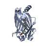
| ||||||||
| 2 | 
| ||||||||
| Unit cell |
|
-Components
| #1: Protein | Mass: 17309.793 Da / Num. of mol.: 2 / Fragment: UNP residues 2-150 Source method: isolated from a genetically manipulated source Source: (gene. exp.) Homo sapiens (human) / Gene: PDE6D, PDED / Production host:  Escherichia coli (E. coli) Escherichia coli (E. coli)References: UniProt: O43924, 3',5'-cyclic-GMP phosphodiesterase#2: Protein/peptide | Mass: 581.726 Da / Num. of mol.: 2 / Fragment: UNP residues 851-855 Source method: isolated from a genetically manipulated source Source: (gene. exp.) Homo sapiens (human) / Gene: PDE6C, PDEA2 / Production host: Escherichia coli (E. coli)References: UniProt: P51160, 3',5'-cyclic-GMP phosphodiesterase#3: Chemical | 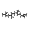  Mass: 274.484 Da / Num. of mol.: 2 Mass: 274.484 Da / Num. of mol.: 2Source method: isolated from a genetically manipulated source Formula: C20H34 / Source: (gene. exp.) Homo sapiens (human) / Production host: Escherichia coli (E. coli)#4: Water | ChemComp-HOH / | Water Mass: 18.015 Da / Num. of mol.: 108 / Source method: isolated from a natural source / Formula: H2O Mass: 18.015 Da / Num. of mol.: 108 / Source method: isolated from a natural source / Formula: H2O |
|---|
-Experimental details
-Experiment
| Experiment | Method: X-RAY DIFFRACTION / Number of used crystals: 1 |
|---|
- Sample preparation
| Crystal | Density Matthews: 2.61 Å3/Da / Density % sol: 52.89 % |
|---|---|
| Crystal grow | Temperature: 293 K / Method: vapor diffusion, sitting drop Details: 0.1 M HEPES (pH 7.5), 0.2 M Li2SO4, 25 % PEG4000 and 0.1 M NaOAc |
-Data collection
| Diffraction | Mean temperature: 100 K |
|---|---|
| Diffraction source | Source: SYNCHROTRON / Site: SLS  / Beamline: X10SA / Wavelength: 1.00001 Å / Beamline: X10SA / Wavelength: 1.00001 Å |
| Detector | Type: DECTRIS PILATUS 6M / Detector: PIXEL / Date: Sep 12, 2014 |
| Radiation | Protocol: SINGLE WAVELENGTH / Monochromatic (M) / Laue (L): M / Scattering type: x-ray |
| Radiation wavelength | Wavelength: 1.00001 Å / Relative weight: 1 |
| Reflection | Resolution: 2.1→29.63 Å / Num. obs: 22232 / % possible obs: 99.6 % / Redundancy: 5.29 % / Rsym value: 0.05 / Net I/σ(I): 23.46 |
| Reflection shell | Resolution: 2.1→2.2 Å / Redundancy: 5.35 % / Mean I/σ(I) obs: 10.24 / Rsym value: 0.163 / % possible all: 100 |
- Processing
| Software |
| |||||||||||||||||||||||||||||||||||||||||||||||||||||||||||||||||||||||||||
|---|---|---|---|---|---|---|---|---|---|---|---|---|---|---|---|---|---|---|---|---|---|---|---|---|---|---|---|---|---|---|---|---|---|---|---|---|---|---|---|---|---|---|---|---|---|---|---|---|---|---|---|---|---|---|---|---|---|---|---|---|---|---|---|---|---|---|---|---|---|---|---|---|---|---|---|---|
| Refinement | Method to determine structure: MOLECULAR REPLACEMENT Starting model: 3T5G Resolution: 2.1→29.63 Å / Cor.coef. Fo:Fc: 0.948 / Cor.coef. Fo:Fc free: 0.913 / SU B: 4.7 / SU ML: 0.129 / Cross valid method: THROUGHOUT / σ(F): 0 / ESU R: 0.212 / ESU R Free: 0.194 / Stereochemistry target values: MAXIMUM LIKELIHOOD Details: HYDROGENS HAVE BEEN ADDED IN THE RIDING POSITIONS U VALUES : REFINED INDIVIDUALLY
| |||||||||||||||||||||||||||||||||||||||||||||||||||||||||||||||||||||||||||
| Solvent computation | Ion probe radii: 0.8 Å / Shrinkage radii: 0.8 Å / VDW probe radii: 1.2 Å / Solvent model: MASK | |||||||||||||||||||||||||||||||||||||||||||||||||||||||||||||||||||||||||||
| Displacement parameters | Biso max: 89.69 Å2 / Biso mean: 34.13 Å2 / Biso min: 17.08 Å2
| |||||||||||||||||||||||||||||||||||||||||||||||||||||||||||||||||||||||||||
| Refinement step | Cycle: final / Resolution: 2.1→29.63 Å
| |||||||||||||||||||||||||||||||||||||||||||||||||||||||||||||||||||||||||||
| Refine LS restraints |
| |||||||||||||||||||||||||||||||||||||||||||||||||||||||||||||||||||||||||||
| LS refinement shell | Resolution: 2.1→2.154 Å / Total num. of bins used: 20
|
