 Movie
Movie Controller

[English] 日本語
 Yorodumi
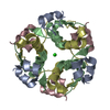
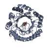
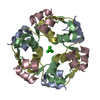
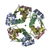
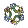
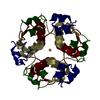
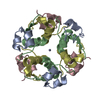
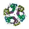
- PDB-5e7w: X-ray Structure of Human Recombinant 2Zn insulin at 0.92 Angstrom -
+ Open data

- Basic information
| Entry | Database: PDB / ID: 5e7w | ||||||
|---|---|---|---|---|---|---|---|
| Title | X-ray Structure of Human Recombinant 2Zn insulin at 0.92 Angstrom | ||||||
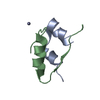
 Components Components | (Insulin ) x 2 ) x 2 | ||||||
 Keywords Keywords | IMMUNE SYSTEM / Insulin / human / recombinant / high-resolution | ||||||
| Function / homology |  Function and homology information Function and homology informationnegative regulation of NAD(P)H oxidase activity / negative regulation of glycogen catabolic process / regulation of cellular amino acid metabolic process / negative regulation of fatty acid metabolic process / negative regulation of feeding behavior / Signaling by Insulin receptor / nitric oxide-cGMP-mediated signaling / IRS activation / Insulin processing / regulation of protein secretion ...negative regulation of NAD(P)H oxidase activity / negative regulation of glycogen catabolic process / regulation of cellular amino acid metabolic process / negative regulation of fatty acid metabolic process / negative regulation of feeding behavior / Signaling by Insulin receptor / nitric oxide-cGMP-mediated signaling / IRS activation / Insulin processing / regulation of protein secretion / positive regulation of peptide hormone secretion / positive regulation of respiratory burst / Regulation of gene expression in beta cells / negative regulation of acute inflammatory response / alpha-beta T cell activation / negative regulation of respiratory burst involved in inflammatory response / positive regulation of dendritic spine maintenance / positive regulation of glycogen biosynthetic process / Synthesis, secretion, and deacylation of Ghrelin / negative regulation of protein secretion / positive regulation of nitric oxide mediated signal transduction / fatty acid homeostasis / regulation of protein localization to plasma membrane / Signal attenuation / FOXO-mediated transcription of oxidative stress, metabolic and neuronal genes / negative regulation of lipid catabolic process / negative regulation of gluconeogenesis / COPI-mediated anterograde transport / positive regulation of lipid biosynthetic process / negative regulation of oxidative stress-induced intrinsic apoptotic signaling pathway / negative regulation of reactive oxygen species biosynthetic process / positive regulation of insulin receptor signaling pathway / transport vesicle / positive regulation of protein autophosphorylation / Insulin receptor recycling / insulin-like growth factor receptor binding / neuron projection maintenance / NPAS4 regulates expression of target genes / positive regulation of protein metabolic process / positive regulation of brown fat cell differentiation / endoplasmic reticulum-Golgi intermediate compartment membrane / positive regulation of glycolytic process / activation of protein kinase B activity / positive regulation of mitotic nuclear division / Insulin receptor signalling cascade / positive regulation of cytokine production / positive regulation of long-term synaptic potentiation / Regulation of insulin secretion / acute-phase response / endosome lumen / positive regulation of protein secretion / positive regulation of glucose import / positive regulation of nitric-oxide synthase activity / positive regulation of cell differentiation / negative regulation of proteolysis / regulation of transmembrane transporter activity / wound healing / regulation of synaptic plasticity / insulin receptor binding / negative regulation of protein catabolic process / hormone activity / cognition / positive regulation of neuron projection development / Golgi lumen / positive regulation of protein localization to nucleus / vasodilation / glucose metabolic process / regulation of protein localization / insulin receptor signaling pathway / cell-cell signaling / glucose homeostasis / positive regulation of NF-kappaB transcription factor activity / PI5P, PP2A and IER3 Regulate PI3K/AKT Signaling / positive regulation of cell growth / secretory granule lumen / protease binding / positive regulation of MAPK cascade / positive regulation of phosphatidylinositol 3-kinase/protein kinase B signal transduction / positive regulation of cell migration / G protein-coupled receptor signaling pathway / Amyloid fiber formation / endoplasmic reticulum lumen / Golgi membrane / negative regulation of gene expression / positive regulation of cell population proliferation / regulation of DNA-templated transcription / positive regulation of gene expression / extracellular space / extracellular region / identical protein bindingSimilarity search - Function | ||||||
| Biological species |  Homo sapiens (human) Homo sapiens (human) | ||||||
| Method | X-RAY DIFFRACTION / SYNCHROTRON / MOLECULAR REPLACEMENT / Resolution: 0.9519 Å | ||||||
 Authors Authors | Lisgarten, D.R. / Naylor, C.E. / Palmer, R.A. / Lobley, C.M.C. | ||||||
 Citation Citation | Journal: Chem Cent J / Year: 2017 Title: Ultra-high resolution X-ray structures of two forms of human recombinant insulin at 100 K. Authors: Lisgarten, D.R. / Palmer, R.A. / Lobley, C.M.C. / Naylor, C.E. / Chowdhry, B.Z. / Al-Kurdi, Z.I. / Badwan, A.A. / Howlin, B.J. / Gibbons, N.C.J. / Saldanha, J.W. / Lisgarten, J.N. / Basak, A.K. | ||||||
| History |
|
- Structure visualization
| Structure viewer | Molecule: MolmilJmol/JSmol |
|---|
- Downloads & links
-Download
| PDBx/mmCIF format | 5e7w.cif.gz | 87.8 KB | Display | PDBx/mmCIF format |
|---|---|---|---|---|
| PDB format | pdb5e7w.ent.gz | 67.9 KB | Display | PDB format |
| PDBx/mmJSON format | 5e7w.json.gz | Tree view | PDBx/mmJSON format | |
| Others |  Other downloads Other downloads |
-Validation report
| Arichive directory | https://data.pdbj.org/pub/pdb/validation_reports/e7/5e7wftp://data.pdbj.org/pub/pdb/validation_reports/e7/5e7w | HTTPS FTP |
|---|
-Related structure data
| Related structure data | 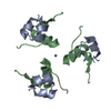 3w7yS S: Starting model for refinement |
|---|---|
| Similar structure data |
-Links
 PDBj
PDBj


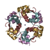
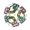
- Assembly
| Deposited unit | 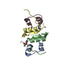
| ||||||||||||||||||
|---|---|---|---|---|---|---|---|---|---|---|---|---|---|---|---|---|---|---|---|
| 1 | 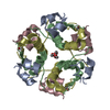
| ||||||||||||||||||
| Unit cell |
| ||||||||||||||||||
| Components on special symmetry positions |
|
-Components
-Protein/peptide , 2 types, 4 molecules ACBD
| #1: Protein/peptide | Mass: 2383.698 Da / Num. of mol.: 2 Source method: isolated from a genetically manipulated source Details: Human Insulin A and C Chain Insulin was purchased from Insugen Source: (gene. exp.) Homo sapiens (human) / Gene: INS / Production host:  Komagataella pastoris (fungus) / References: UniProt: P01308 Komagataella pastoris (fungus) / References: UniProt: P01308#2: Protein/peptide | Mass: 3433.953 Da / Num. of mol.: 2 Source method: isolated from a genetically manipulated source Details: Human Insulin B and D chain Insulin was purchased from Insugen Source: (gene. exp.) Homo sapiens (human) / Gene: INS / Production host: Komagataella pastoris (fungus) / References: UniProt: P01308 |
|---|
-Non-polymers , 4 types, 224 molecules 


| #3: Chemical |  Mass: 65.409 Da / Num. of mol.: 2 / Source method: obtained synthetically / Formula: Zn Mass: 65.409 Da / Num. of mol.: 2 / Source method: obtained synthetically / Formula: Zn#4: Chemical | ChemComp-ACT / | Acetate Mass: 59.044 Da / Num. of mol.: 1 / Source method: obtained synthetically / Formula: C2H3O2 Mass: 59.044 Da / Num. of mol.: 1 / Source method: obtained synthetically / Formula: C2H3O2#5: Chemical | ChemComp-POL / | Propan-1-ol Mass: 60.095 Da / Num. of mol.: 1 / Source method: obtained synthetically / Formula: C3H8O Mass: 60.095 Da / Num. of mol.: 1 / Source method: obtained synthetically / Formula: C3H8O#6: Water | ChemComp-HOH / | WaterMass: 18.015 Da / Num. of mol.: 220 / Source method: isolated from a natural source / Formula: H2O |
|---|
-Experimental details
-Experiment
| Experiment | Method: X-RAY DIFFRACTION |
|---|
- Sample preparation
| Crystal | Density Matthews: 1.89 Å3/Da / Density % sol: 35 % / Description: needle |
|---|---|
| Crystal grow | Temperature: 293 K / Method: batch mode / pH: 6.3 Details: The crystals were prepared by a batch method similar to that of Baker et al, 1988 [1], modified as follows: 0.01g of insulin as a fine powder was placed in a clean test tube; 0.02M HCl was ...Details: The crystals were prepared by a batch method similar to that of Baker et al, 1988 [1], modified as follows: 0.01g of insulin as a fine powder was placed in a clean test tube; 0.02M HCl was added to dissolve the protein; on addition of 0.15 mL of 0.15 M zinc acetate the solution became cloudy due to precipitation of the protein; 0.3 mL of acetone and then 0.5 mL of trisodium citrate together with 0.8 mL of water were added and the solution went clear; the pH was checked and increased with NaOH to a pH between 8 and 9 for different batches, thus ensuring complete dissolution. It was then adjusted to the required value of pH 6.3. If any slight turbidity occurred, it was removed by warming the solution. The solution was then filtered using a Millipore membrane/acetate cellulose acetate filter. This removes any nuclei which will encourage precipitation or formation of masses of small crystals. The solution was then warmed to 50 deg C by surrounding the test tube with preheated water in a Dewar. This allowed the solution to cool slowly to room temperature. The test tube was lightly sealed with cling film; crystals formed within a few days and were of suitable size for X-ray diffraction within two weeks; the test tube containing crystals was kept at 4 degC prior to data collection. The crystal used for data collection was about 0.2 mm3. PH range: 6.2 - 6.4 / Temp details: Room Temperature |
-Data collection
| Diffraction | Mean temperature: 100 K |
|---|---|
| Diffraction source | Source: SYNCHROTRON / Site: Diamond  / Beamline: I02 / Wavelength: 0.77 Å / Beamline: I02 / Wavelength: 0.77 Å |
| Detector | Type: DECTRIS PILATUS 6M / Detector: PIXEL / Date: Sep 27, 2012 Details: Data were collected at 16000keV (0.77 Angstrom) and 100 deg K with the Pilatus 6M detector as close to the sample as possible (179.5mm). The EDNA strategy was used to obtain a start angle ...Details: Data were collected at 16000keV (0.77 Angstrom) and 100 deg K with the Pilatus 6M detector as close to the sample as possible (179.5mm). The EDNA strategy was used to obtain a start angle and 180 deg of data were collected with 0.1 deg oscillation and 0.1s exposure. The resolution of useful diffraction data achieved and used for structure analysis was 0.92 Angstrom. |
| Radiation | Monochromator: Beamline fixed at 16000keV / Protocol: SINGLE WAVELENGTH / Monochromatic (M) / Laue (L): M / Scattering type: x-ray |
| Radiation wavelength | Wavelength: 0.77 Å / Relative weight: 1 |
| Reflection | Resolution: 0.92→40.91 Å / Num. obs: 58647 / % possible obs: 100 % / Redundancy: 5 % / Rmerge(I) obs: 0.034 / Net I/σ(I): 20.2 |
| Reflection shell | Resolution: 0.92→0.94 Å / Redundancy: 4.7 % / Rmerge(I) obs: 0.967 / Mean I/σ(I) obs: 1.6 / % possible all: 100 |
- Processing
| Software |
| ||||||||||||||||||||
|---|---|---|---|---|---|---|---|---|---|---|---|---|---|---|---|---|---|---|---|---|---|
| Refinement | Method to determine structure: MOLECULAR REPLACEMENT Starting model: 3W7Y Resolution: 0.9519→10 Å / Cross valid method: FREE R-VALUE / σ(F): 0
| ||||||||||||||||||||
| Displacement parameters | Biso mean: 18.45 Å2 | ||||||||||||||||||||
| Refinement step | Cycle: LAST / Resolution: 0.9519→10 Å
|
