 Movie
Movie Controller


+ Open data

- Basic information
| Entry | Database: PDB / ID: 5.0E+44 | ||||||
|---|---|---|---|---|---|---|---|
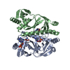
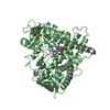
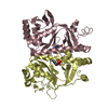
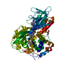
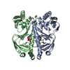
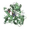
| Title | Crystal structure of holo-FNR of A. fischeri | ||||||
 Components Components | FNR regulator | ||||||
 Keywords Keywords |  TRANSCRIPTION / labile dimer TRANSCRIPTION / labile dimer | ||||||
| Function / homology |  Function and homology information Function and homology information2 iron, 2 sulfur cluster binding / 4 iron, 4 sulfur cluster binding / DNA-binding transcription factor activity / DNA binding / metal ion bindingSimilarity search - Function | ||||||
| Biological species |  Aliivibrio fischeri (bacteria) Aliivibrio fischeri (bacteria) | ||||||
| Method | X-RAY DIFFRACTION / SYNCHROTRON / MOLECULAR REPLACEMENT / Resolution: 2.65 Å | ||||||
 Authors Authors | Volbeda, A. / Fontecilla-Camps, J.C. | ||||||
 Citation Citation | Journal: Sci Adv / Year: 2015 Title: The crystal structure of the global anaerobic transcriptional regulator FNR explains its extremely fine-tuned monomer-dimer equilibrium. Authors: Volbeda, A. / Darnault, C. / Renoux, O. / Nicolet, Y. / Fontecilla-Camps, J.C. | ||||||
| History |
|
- Structure visualization
| Structure viewer | Molecule: MolmilJmol/JSmol |
|---|
- Downloads & links
-Download
| PDBx/mmCIF format | 5e44.cif.gz | 107.9 KB | Display | PDBx/mmCIF format |
|---|---|---|---|---|
| PDB format | pdb5e44.ent.gz | 82.3 KB | Display | PDB format |
| PDBx/mmJSON format | 5e44.json.gz | Tree view | PDBx/mmJSON format | |
| Others |  Other downloads Other downloads |
-Validation report
| Arichive directory | https://data.pdbj.org/pub/pdb/validation_reports/e4/5e44ftp://data.pdbj.org/pub/pdb/validation_reports/e4/5e44 | HTTPS FTP |
|---|
-Related structure data
| Related structure data | 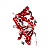 5cvrC 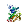 4i20S S: Starting model for refinement C: citing same article ( |
|---|---|
| Similar structure data |


-Links
 PDBj
PDBj


- Assembly
| Deposited unit | 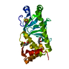
| ||||||||
|---|---|---|---|---|---|---|---|---|---|
| 1 | 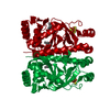
| ||||||||
| Unit cell |
|
-Components
| #1: Protein | Mass: 29223.398 Da / Num. of mol.: 1 / Fragment: UNP residues 3-250 Source method: isolated from a genetically manipulated source Source: (gene. exp.) Aliivibrio fischeri (bacteria) / Gene: fnr / Plasmid: pET-15b / Production host: Escherichia coli BL21(DE3) (bacteria) / References: UniProt: Q70ET4 |
|---|---|
| #2: Chemical | ChemComp-SF4 / Iron–sulfur cluster  Mass: 351.640 Da / Num. of mol.: 1 / Source method: obtained synthetically / Formula: Fe4S4 Mass: 351.640 Da / Num. of mol.: 1 / Source method: obtained synthetically / Formula: Fe4S4 |
| #3: Chemical | 2-Methyl-2,4-pentanediol  Mass: 118.174 Da / Num. of mol.: 2 / Source method: obtained synthetically / Formula: C6H14O2 / Comment: precipitant*YM Mass: 118.174 Da / Num. of mol.: 2 / Source method: obtained synthetically / Formula: C6H14O2 / Comment: precipitant*YM |
-Experimental details
-Experiment
| Experiment | Method: X-RAY DIFFRACTION / Number of used crystals: 1 |
|---|
- Sample preparation
| Crystal | Density Matthews: 2.66 Å3/Da / Density % sol: 53.74 % |
|---|---|
| Crystal grow | Temperature: 298 K / Method: vapor diffusion / pH: 6.4 / Details: MPD, MES, dithionite, anaerobic |
-Data collection
| Diffraction | Mean temperature: 100 K | |||||||||||||||||||||||||||
|---|---|---|---|---|---|---|---|---|---|---|---|---|---|---|---|---|---|---|---|---|---|---|---|---|---|---|---|---|
| Diffraction source | Source: SYNCHROTRON / Site: ESRF  / Beamline: ID23-2 / Wavelength: 0.8726 Å / Beamline: ID23-2 / Wavelength: 0.8726 Å | |||||||||||||||||||||||||||
| Detector | Type: DECTRIS PILATUS 6M-F / Detector: PIXEL / Date: May 4, 2015 | |||||||||||||||||||||||||||
| Radiation | Protocol: SINGLE WAVELENGTH / Monochromatic (M) / Laue (L): M / Scattering type: x-ray | |||||||||||||||||||||||||||
| Radiation wavelength | Wavelength: 0.8726 Å / Relative weight: 1 | |||||||||||||||||||||||||||
| Reflection | Resolution: 2.65→47.95 Å / Num. all: 9630 / Num. obs: 9607 / % possible obs: 99.78 % / Redundancy: 25.4 % / Biso Wilson estimate: 82.7 Å2 / CC1/2: 0.997 / Rmerge(I) obs: 0.172 / Rpim(I) all: 0.036 / Net I/σ(I): 14.2 / Num. measured all: 244255 / Scaling rejects: 6 | |||||||||||||||||||||||||||
| Reflection shell | Diffraction-ID: 1 / Rejects: 0
|
- Processing
| Software |
| |||||||||||||||||||||||||||||||||||||||||||||||||||||||||||||||||||||||||||||||||||||||||||||||||||||||||||||||||||||||||||||
|---|---|---|---|---|---|---|---|---|---|---|---|---|---|---|---|---|---|---|---|---|---|---|---|---|---|---|---|---|---|---|---|---|---|---|---|---|---|---|---|---|---|---|---|---|---|---|---|---|---|---|---|---|---|---|---|---|---|---|---|---|---|---|---|---|---|---|---|---|---|---|---|---|---|---|---|---|---|---|---|---|---|---|---|---|---|---|---|---|---|---|---|---|---|---|---|---|---|---|---|---|---|---|---|---|---|---|---|---|---|---|---|---|---|---|---|---|---|---|---|---|---|---|---|---|---|---|
| Refinement | Method to determine structure: MOLECULAR REPLACEMENT Starting model: 4i20 Resolution: 2.65→47.949 Å / SU ML: 0.45 / Cross valid method: THROUGHOUT / σ(F): 1.34 / Phase error: 27.08 / Stereochemistry target values: ML Details: BIJVOET PAIRS WERE TREATED AS INDEPENDENT OBSERVATIONS ALLOWING TO MODEL ANOMALOUS CONTRIBUTIONS OF FE AND S ATOMS, AS THIS GAVE SIGNIFICANTLY IMPROVED REFINEMENT STATISTICS
| |||||||||||||||||||||||||||||||||||||||||||||||||||||||||||||||||||||||||||||||||||||||||||||||||||||||||||||||||||||||||||||
| Solvent computation | Shrinkage radii: 0.9 Å / VDW probe radii: 1.11 Å / Solvent model: FLAT BULK SOLVENT MODEL | |||||||||||||||||||||||||||||||||||||||||||||||||||||||||||||||||||||||||||||||||||||||||||||||||||||||||||||||||||||||||||||
| Displacement parameters | Biso max: 178.32 Å2 / Biso mean: 96.0049 Å2 / Biso min: 53.41 Å2 | |||||||||||||||||||||||||||||||||||||||||||||||||||||||||||||||||||||||||||||||||||||||||||||||||||||||||||||||||||||||||||||
| Refinement step | Cycle: final / Resolution: 2.65→47.949 Å
| |||||||||||||||||||||||||||||||||||||||||||||||||||||||||||||||||||||||||||||||||||||||||||||||||||||||||||||||||||||||||||||
| Refine LS restraints |
| |||||||||||||||||||||||||||||||||||||||||||||||||||||||||||||||||||||||||||||||||||||||||||||||||||||||||||||||||||||||||||||
| LS refinement shell | Refine-ID: X-RAY DIFFRACTION / Total num. of bins used: 5
| |||||||||||||||||||||||||||||||||||||||||||||||||||||||||||||||||||||||||||||||||||||||||||||||||||||||||||||||||||||||||||||
| Refinement TLS params. | Method: refined / Refine-ID: X-RAY DIFFRACTION
| |||||||||||||||||||||||||||||||||||||||||||||||||||||||||||||||||||||||||||||||||||||||||||||||||||||||||||||||||||||||||||||
| Refinement TLS group |
|
