 Movie
Movie Controller


+ Open data

- Basic information
| Entry | Database: PDB / ID: 4ivo | ||||||
|---|---|---|---|---|---|---|---|
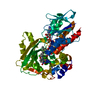
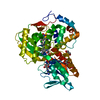
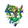
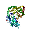
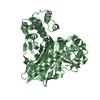
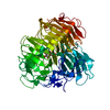
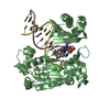
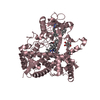
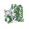
| Title | Structure of human protoporphyrinogen IX oxidase(R59Q) | ||||||
 Components Components | Protoporphyrinogen oxidase | ||||||
 Keywords Keywords | OXIDOREDUCTASE / Oxidase / FAD Binding / Membrane | ||||||
| Function / homology |  Function and homology information Function and homology informationphytoene dehydrogenase activity / protoporphyrinogen oxidase / oxygen-dependent protoporphyrinogen oxidase activity / carotenoid biosynthetic process / heme B biosynthetic process / heme O biosynthetic process / heme A biosynthetic process / porphyrin-containing compound biosynthetic process / protoporphyrinogen IX biosynthetic process / Heme biosynthesis ...phytoene dehydrogenase activity / protoporphyrinogen oxidase / oxygen-dependent protoporphyrinogen oxidase activity / carotenoid biosynthetic process / heme B biosynthetic process / heme O biosynthetic process / heme A biosynthetic process / porphyrin-containing compound biosynthetic process / protoporphyrinogen IX biosynthetic process / Heme biosynthesis / heme biosynthetic process / mitochondrial membrane / mitochondrial intermembrane space / flavin adenine dinucleotide binding / mitochondrial inner membraneSimilarity search - Function | ||||||
| Biological species |  Homo sapiens (human) Homo sapiens (human) | ||||||
| Method | X-RAY DIFFRACTION / SYNCHROTRON / MOLECULAR REPLACEMENT / Resolution: 2.597 Å | ||||||
 Authors Authors | Xiaohong, Q. / Baifan, W. | ||||||
 Citation Citation | Journal: J.Biol.Chem. / Year: 2013 Title: Quantitative structural insight into human variegate porphyria disease. Authors: Wang, B. / Wen, X. / Qin, X. / Wang, Z. / Tan, Y. / Shen, Y. / Xi, Z. | ||||||
| History |
|
- Structure visualization
| Structure viewer | Molecule: MolmilJmol/JSmol |
|---|
- Downloads & links
-Download
| PDBx/mmCIF format | 4ivo.cif.gz | 104.9 KB | Display | PDBx/mmCIF format |
|---|---|---|---|---|
| PDB format | pdb4ivo.ent.gz | 78.6 KB | Display | PDB format |
| PDBx/mmJSON format | 4ivo.json.gz | Tree view | PDBx/mmJSON format | |
| Others |  Other downloads Other downloads |
-Validation report
| Arichive directory | https://data.pdbj.org/pub/pdb/validation_reports/iv/4ivoftp://data.pdbj.org/pub/pdb/validation_reports/iv/4ivo | HTTPS FTP |
|---|
-Related structure data


-Links
 PDBj
PDBj


- Assembly
| Deposited unit | 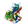
| ||||||||
|---|---|---|---|---|---|---|---|---|---|
| 1 |
| ||||||||
| 2 | 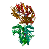
| ||||||||
| Unit cell |
|
-Components
| #1: Protein | / PPO Mass: 51620.918 Da / Num. of mol.: 1 / Mutation: R59Q Source method: isolated from a genetically manipulated source Source: (gene. exp.) Homo sapiens (human) / Gene: PPOX / Plasmid: pTrcHis B / Production host:  Escherichia coli (E. coli) / Strain (production host): BL21(DE3)pLysS / References: UniProt: P50336, protoporphyrinogen oxidase Escherichia coli (E. coli) / Strain (production host): BL21(DE3)pLysS / References: UniProt: P50336, protoporphyrinogen oxidase | ||
|---|---|---|---|
| #2: Chemical | ChemComp-ACJ / Acifluorfen  Mass: 361.657 Da / Num. of mol.: 1 / Source method: obtained synthetically / Formula: C14H7ClF3NO5 Mass: 361.657 Da / Num. of mol.: 1 / Source method: obtained synthetically / Formula: C14H7ClF3NO5 | ||
| #3: Chemical | ChemComp-FAD / Flavin adenine dinucleotide  Mass: 785.550 Da / Num. of mol.: 1 / Source method: obtained synthetically / Formula: C27H33N9O15P2 / Comment: FAD*YM Mass: 785.550 Da / Num. of mol.: 1 / Source method: obtained synthetically / Formula: C27H33N9O15P2 / Comment: FAD*YM | ||
| #4: Chemical | Glycerol  Mass: 92.094 Da / Num. of mol.: 2 / Source method: obtained synthetically / Formula: C3H8O3 Mass: 92.094 Da / Num. of mol.: 2 / Source method: obtained synthetically / Formula: C3H8O3#5: Water | ChemComp-HOH / | Water Mass: 18.015 Da / Num. of mol.: 76 / Source method: isolated from a natural source / Formula: H2O Mass: 18.015 Da / Num. of mol.: 76 / Source method: isolated from a natural source / Formula: H2O |
-Experimental details
-Experiment
| Experiment | Method: X-RAY DIFFRACTION / Number of used crystals: 1 |
|---|
- Sample preparation
| Crystal | Density Matthews: 2.72 Å3/Da / Density % sol: 54.81 % |
|---|---|
| Crystal grow | Temperature: 298 K / Method: vapor diffusion, sitting drop / pH: 10 Details: ammonium citrate dibasic, PEG3350, pH 10.0, VAPOR DIFFUSION, SITTING DROP, temperature 298K |
-Data collection
| Diffraction | Mean temperature: 100 K |
|---|---|
| Diffraction source | Source: SYNCHROTRON / Site: SSRF  / Beamline: BL17U / Wavelength: 0.9796 Å / Beamline: BL17U / Wavelength: 0.9796 Å |
| Detector | Type: ADSC QUANTUM 210 / Detector: CCD / Date: Jun 2, 2010 |
| Radiation | Monochromator: GRAPHITE / Protocol: SINGLE WAVELENGTH / Monochromatic (M) / Laue (L): M / Scattering type: x-ray |
| Radiation wavelength | Wavelength: 0.9796 Å / Relative weight: 1 |
| Reflection | Resolution: 2.6→50 Å / Num. all: 17528 / Num. obs: 16944 / % possible obs: 99.9 % / Observed criterion σ(F): 2 / Observed criterion σ(I): 2 |
| Reflection shell | Resolution: 2.6→2.69 Å / % possible all: 99.7 |
- Processing
| Software |
| |||||||||||||||||||||||||||||||||||||||||||||||||
|---|---|---|---|---|---|---|---|---|---|---|---|---|---|---|---|---|---|---|---|---|---|---|---|---|---|---|---|---|---|---|---|---|---|---|---|---|---|---|---|---|---|---|---|---|---|---|---|---|---|---|
| Refinement | Method to determine structure: MOLECULAR REPLACEMENT / Resolution: 2.597→30.579 Å / SU ML: 0.31 / σ(F): 0 / Phase error: 21.92 / Stereochemistry target values: ML
| |||||||||||||||||||||||||||||||||||||||||||||||||
| Solvent computation | Shrinkage radii: 0.72 Å / VDW probe radii: 1 Å / Solvent model: FLAT BULK SOLVENT MODEL / Bsol: 20.46 Å2 / ksol: 0.351 e/Å3 | |||||||||||||||||||||||||||||||||||||||||||||||||
| Displacement parameters |
| |||||||||||||||||||||||||||||||||||||||||||||||||
| Refinement step | Cycle: LAST / Resolution: 2.597→30.579 Å
| |||||||||||||||||||||||||||||||||||||||||||||||||
| Refine LS restraints |
| |||||||||||||||||||||||||||||||||||||||||||||||||
| LS refinement shell |
|
