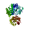
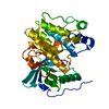
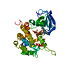
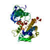
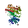
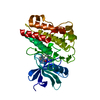
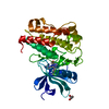
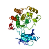
[English] 日本語
Yorodumi
- PDB-5e1s: The Crystal structure of INSR Tyrosine Kinase in complex with the... -

+
Open data


ID or keywords:

Loading...

-
Basic information

Entry
Database: PDB / ID: 5e1s
TitleThe Crystal structure of INSR Tyrosine Kinase in complex with the Inhibitor BI 885578
ComponentsInsulin receptor
KeywordsTRANSFERASE / Kinase / INSR / IGF-1R / Inhibitor
Function / homology
Function and homology information


regulation of female gonad development / positive regulation of meiotic cell cycle / positive regulation of developmental growth / insulin-like growth factor II binding / male sex determination / exocrine pancreas development / insulin receptor complex / insulin-like growth factor I binding / insulin receptor activity / positive regulation of protein-containing complex disassembly ...regulation of female gonad development / positive regulation of meiotic cell cycle / positive regulation of developmental growth / insulin-like growth factor II binding / male sex determination / exocrine pancreas development / insulin receptor complex / insulin-like growth factor I binding / insulin receptor activity / positive regulation of protein-containing complex disassembly / cargo receptor activity / dendritic spine maintenance / insulin binding / PTB domain binding / adrenal gland development / neuronal cell body membrane / Signaling by Insulin receptor / IRS activation / activation of protein kinase activity / amyloid-beta clearance / positive regulation of respiratory burst / regulation of embryonic development / positive regulation of receptor internalization / transport across blood-brain barrier / insulin receptor substrate binding / positive regulation of glycogen biosynthetic process / epidermis development / Signal attenuation / phosphatidylinositol 3-kinase binding / heart morphogenesis / dendrite membrane / Insulin receptor recycling / insulin-like growth factor receptor binding / neuron projection maintenance / positive regulation of glycolytic process / activation of protein kinase B activity / positive regulation of mitotic nuclear division / Insulin receptor signalling cascade / receptor-mediated endocytosis / learning / caveola / positive regulation of glucose import / positive regulation of MAP kinase activity / receptor internalization / receptor protein-tyrosine kinase / memory / cellular response to growth factor stimulus / peptidyl-tyrosine phosphorylation / cellular response to insulin stimulus / positive regulation of nitric oxide biosynthetic process / male gonad development / late endosome / insulin receptor signaling pathway / glucose homeostasis / amyloid-beta binding / PI5P, PP2A and IER3 Regulate PI3K/AKT Signaling / protein tyrosine kinase activity / positive regulation of MAPK cascade / protein autophosphorylation / positive regulation of phosphatidylinositol 3-kinase/protein kinase B signal transduction / lysosome / receptor complex / endosome membrane / positive regulation of cell migration / symbiont entry into host cell / positive regulation of protein phosphorylation / G protein-coupled receptor signaling pathway / protein domain specific binding / axon / external side of plasma membrane / protein phosphorylation / positive regulation of cell population proliferation / protein-containing complex binding / regulation of DNA-templated transcription / GTP binding / positive regulation of DNA-templated transcription / extracellular exosome / ATP binding / membrane / identical protein binding / plasma membrane
Similarity search - Function
Insulin receptor, trans-membrane domain / Insulin receptor trans-membrane segment / Tyrosine-protein kinase, insulin-like receptor / Tyrosine-protein kinase, receptor class II, conserved site / Receptor tyrosine kinase class II signature. / Receptor L-domain / Furin-like cysteine-rich domain / Receptor L-domain superfamily / Furin-like cysteine rich region / Receptor L domain ...Insulin receptor, trans-membrane domain / Insulin receptor trans-membrane segment / Tyrosine-protein kinase, insulin-like receptor / Tyrosine-protein kinase, receptor class II, conserved site / Receptor tyrosine kinase class II signature. / Receptor L-domain / Furin-like cysteine-rich domain / Receptor L-domain superfamily / Furin-like cysteine rich region / Receptor L domain / Furin-like repeat / Furin-like repeats / Growth factor receptor cysteine-rich domain superfamily / Fibronectin type III domain / Fibronectin type 3 domain / Fibronectin type-III domain profile. / Fibronectin type III / Fibronectin type III superfamily / Tyrosine-protein kinase, catalytic domain / Tyrosine kinase, catalytic domain / Tyrosine protein kinases specific active-site signature. / Tyrosine-protein kinase, active site / Protein tyrosine and serine/threonine kinase / Serine-threonine/tyrosine-protein kinase, catalytic domain / Transferase(Phosphotransferase) domain 1 / Transferase(Phosphotransferase); domain 1 / Phosphorylase Kinase; domain 1 / Phosphorylase Kinase; domain 1 / Protein kinase, ATP binding site / Protein kinases ATP-binding region signature. / Immunoglobulin-like fold / Protein kinase domain profile. / Protein kinase domain / Protein kinase-like domain superfamily / 2-Layer Sandwich / Orthogonal Bundle / Mainly Alpha / Alpha Beta
Similarity search - Domain/homology
Chem-5JA / Insulin receptor
Similarity search - Component
Biological speciesHomo sapiens (human)
MethodX-RAY DIFFRACTION / SYNCHROTRON / MOLECULAR REPLACEMENT / Resolution: 2.264 Å
AuthorsKessler, D. / Zahn, S. / Sanderson, M. / Wolkerstorfer, B.
CitationJournal: Mol.Cancer Ther. / Year: 2015
Title: BI 885578, a Novel IGF1R/INSR Tyrosine Kinase Inhibitor with Pharmacokinetic Properties That Dissociate Antitumor Efficacy and Perturbation of Glucose Homeostasis.
Authors: Sanderson, M.P. / Apgar, J. / Garin-Chesa, P. / Hofmann, M.H. / Kessler, D. / Quant, J. / Savchenko, A. / Schaaf, O. / Treu, M. / Tye, H. / Zahn, S.K. / Zoephel, A. / Haaksma, E. / Adolf, G.R. / Kraut, N.
History
DepositionSep 30, 2015Deposition site: RCSB / Processing site: PDBE
Revision 1.0Oct 14, 2015Provider: repository / Type: Initial release
Revision 1.1Dec 16, 2015Group: Database references
Revision 1.2Jan 10, 2024Group: Data collection / Database references / Refinement description
Category: chem_comp_atom / chem_comp_bond ...chem_comp_atom / chem_comp_bond / database_2 / pdbx_initial_refinement_model
Item: _database_2.pdbx_DOI / _database_2.pdbx_database_accession

-
Structure visualization

Structure viewerMolecule:
MolmilJmol/JSmol

Downloads & links

-
Assembly

Deposited unit
A: Insulin receptor
hetero molecules


Theoretical massNumber of molelcules
Total (without water)35,4802
Polymers34,9521
Non-polymers5281
Water2,468137
1


  • Idetical with deposited unit
  • defined by software
TypeNameSymmetry operationNumber
identity operation1_555x,y,z1
Buried area0 Å2
ΔGint0 kcal/mol
Surface area14560 Å2
MethodPISA
Unit cell
Length a, b, c (Å)56.550, 70.370, 87.850
Angle α, β, γ (deg.)90.00, 90.00, 90.00
Int Tables number19
Space group name H-MP212121

-
Components

#1: Protein Insulin receptor / / IR


Mass: 34951.949 Da / Num. of mol.: 1
Source method: isolated from a genetically manipulated source
Source: (gene. exp.) Homo sapiens (human) / Gene: INSR / Production host: Spodoptera frugiperda (fall armyworm)
References: UniProt: P06213, receptor protein-tyrosine kinase
#2: Chemical ChemComp-5JA / (5R)-N-(1-{2-[4-(2-methoxyethyl)piperazin-1-yl]ethyl}-1H-pyrazol-3-yl)-5,8-dimethyl-9-phenyl-6,8-dihydro-5H-pyrazolo[3,4-h]quinazolin-2-amine


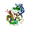
Mass: 527.664 Da / Num. of mol.: 1 / Source method: obtained synthetically / Formula: C29H37N9O
#3: Water ChemComp-HOH / water / Water


Mass: 18.015 Da / Num. of mol.: 137 / Source method: isolated from a natural source / Formula: H2O

-
Experimental details

-
Experiment

ExperimentMethod: X-RAY DIFFRACTION

-
Sample preparation

CrystalDensity Matthews: 2.5 Å3/Da / Density % sol: 50.81 %
Crystal growTemperature: 293 K / Method: vapor diffusion, hanging drop / pH: 7.3
Details: 25% PEG 8000, 0.1M TRIS pH 7.3, 50mM NaCl, 5 mM DTT

-
Data collection

DiffractionMean temperature: 100 K
Diffraction sourceSource: SYNCHROTRON / Site: SLS / Beamline: X06SA / Wavelength: 1 Å
DetectorType: DECTRIS PILATUS 6M-F / Detector: PIXEL / Date: Mar 1, 2013
RadiationProtocol: SINGLE WAVELENGTH / Monochromatic (M) / Laue (L): M / Scattering type: x-ray
Radiation wavelengthWavelength: 1 Å / Relative weight: 1
ReflectionResolution: 2.264→37.26 Å / Num. all: 16922 / Num. obs: 16922 / % possible obs: 100 % / Redundancy: 6.4 % / Biso Wilson estimate: 46.57 Å2 / Rmerge(I) obs: 0.085 / Rsym value: 0.085 / Net I/av σ(I): 14.3 / Net I/σ(I): 14.3
Reflection shellResolution: 2.264→2.272 Å / Redundancy: 6.6 % / Rmerge(I) obs: 0.636 / Mean I/σ(I) obs: 3 / % possible all: 100

-
Processing

Software
NameVersionClassification
BUSTER2.11.2refinement
XDSdata reduction
SCALAdata scaling
PHASERphasing
Cootmodel building
RefinementMethod to determine structure: MOLECULAR REPLACEMENT
Starting model: 1IRK
Resolution: 2.264→37.26 Å / Cor.coef. Fo:Fc: 0.9234 / Cor.coef. Fo:Fc free: 0.8922 / SU R Cruickshank DPI: 0.282 / Cross valid method: THROUGHOUT / σ(F): 0 / SU R Blow DPI: 0.302 / SU Rfree Blow DPI: 0.229 / SU Rfree Cruickshank DPI: 0.225
RfactorNum. reflection% reflectionSelection details
Rfree0.262 851 5.04 %RANDOM
Rwork0.2126 ---
obs0.215 16877 99.38 %-
Displacement parametersBiso mean: 48.74 Å2
Baniso -1Baniso -2Baniso -3
1-7.3803 Å20 Å20 Å2
2---6.581 Å20 Å2
3----0.7993 Å2
Refine analyzeLuzzati coordinate error obs: 0.331 Å
Refinement stepCycle: LAST / Resolution: 2.264→37.26 Å
ProteinNucleic acidLigandSolventTotal
Num. atoms2269 0 39 137 2445
Refine LS restraints
Refine-IDTypeDev idealNumberRestraint functionWeight
X-RAY DIFFRACTIONt_bond_d0.0082366HARMONIC2
X-RAY DIFFRACTIONt_angle_deg0.983204HARMONIC2
X-RAY DIFFRACTIONt_dihedral_angle_d838SINUSOIDAL2
X-RAY DIFFRACTIONt_incorr_chiral_ct
X-RAY DIFFRACTIONt_pseud_angle
X-RAY DIFFRACTIONt_trig_c_planes61HARMONIC2
X-RAY DIFFRACTIONt_gen_planes336HARMONIC5
X-RAY DIFFRACTIONt_it2366HARMONIC20
X-RAY DIFFRACTIONt_nbd
X-RAY DIFFRACTIONt_omega_torsion2.54
X-RAY DIFFRACTIONt_other_torsion16.76
X-RAY DIFFRACTIONt_improper_torsion
X-RAY DIFFRACTIONt_chiral_improper_torsion289SEMIHARMONIC5
X-RAY DIFFRACTIONt_sum_occupancies1HARMONIC1
X-RAY DIFFRACTIONt_utility_distance
X-RAY DIFFRACTIONt_utility_angle
X-RAY DIFFRACTIONt_utility_torsion
X-RAY DIFFRACTIONt_ideal_dist_contact2743SEMIHARMONIC4
LS refinement shellResolution: 2.26→2.42 Å / Total num. of bins used: 8
RfactorNum. reflection% reflection
Rfree0.2507 160 5.5 %
Rwork0.2276 2747 -
all0.2289 2907 -
obs--99.38 %
Refinement TLS params.Method: refined / Origin x: 13.7278 Å / Origin y: 14.502 Å / Origin z: 17.8066 Å
111213212223313233
T-0.1814 Å2-0.0058 Å20.0154 Å2--0.1119 Å2-0.0089 Å2---0.1278 Å2
L1.9788 °2-1.0229 °2-0.1155 °2-1.891 °20.2039 °2--2.1436 °2
S-0.0586 Å °-0.0461 Å °0.1267 Å °0.0671 Å °0.0645 Å °-0.1357 Å °-0.0266 Å °-0.1118 Å °-0.0059 Å °
Refinement TLS groupSelection details: { A|* }

+
About Yorodumi

-
News

-
Feb 9, 2022. New format data for meta-information of EMDB entries

New format data for meta-information of EMDB entries

  • Version 3 of the EMDB header file is now the official format.
  • The previous official version 1.9 will be removed from the archive.

Related info.:EMDB header

External links:wwPDB to switch to version 3 of the EMDB data model

-
Aug 12, 2020. Covid-19 info

Covid-19 info

URL: https://pdbj.org/emnavi/covid19.php

New page: Covid-19 featured information page in EM Navigator.

Related info.:Covid-19 info / Mar 5, 2020. Novel coronavirus structure data

+
Mar 5, 2020. Novel coronavirus structure data

Novel coronavirus structure data

Related info.:Yorodumi Speices / Aug 12, 2020. Covid-19 info

External links:COVID-19 featured content - PDBj / Molecule of the Month (242):Coronavirus Proteases

+
Jan 31, 2019. EMDB accession codes are about to change! (news from PDBe EMDB page)

EMDB accession codes are about to change! (news from PDBe EMDB page)

  • The allocation of 4 digits for EMDB accession codes will soon come to an end. Whilst these codes will remain in use, new EMDB accession codes will include an additional digit and will expand incrementally as the available range of codes is exhausted. The current 4-digit format prefixed with “EMD-” (i.e. EMD-XXXX) will advance to a 5-digit format (i.e. EMD-XXXXX), and so on. It is currently estimated that the 4-digit codes will be depleted around Spring 2019, at which point the 5-digit format will come into force.
  • The EM Navigator/Yorodumi systems omit the EMD- prefix.

Related info.:Q: What is EMD? / ID/Accession-code notation in Yorodumi/EM Navigator

External links:EMDB Accession Codes are Changing Soon! / Contact to PDBj

+
Jul 12, 2017. Major update of PDB

Major update of PDB

  • wwPDB released updated PDB data conforming to the new PDBx/mmCIF dictionary.
  • This is a major update changing the version number from 4 to 5, and with Remediation, in which all the entries are updated.
  • In this update, many items about electron microscopy experimental information are reorganized (e.g. em_software).
  • Now, EM Navigator and Yorodumi are based on the updated data.

External links:wwPDB Remediation / Enriched Model Files Conforming to OneDep Data Standards Now Available in the PDB FTP Archive

-
Yorodumi

Thousand views of thousand structures

  • Yorodumi is a browser for structure data from EMDB, PDB, SASBDB, etc.
  • This page is also the successor to EM Navigator detail page, and also detail information page/front-end page for Omokage search.
  • The word "yorodu" (or yorozu) is an old Japanese word meaning "ten thousand". "mi" (miru) is to see.

Related info.:EMDB / PDB / SASBDB / Comparison of 3 databanks / Yorodumi Search / Aug 31, 2016. New EM Navigator & Yorodumi / Yorodumi Papers / Jmol/JSmol / Function and homology information / Changes in new EM Navigator and Yorodumi

Read more