| Entry | Database: PDB / ID: 5dnx
|
|---|
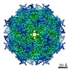
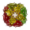
| Title | Crystal structure of IGPD from Pyrococcus furiosus in complex with (R)-C348 |
|---|
 Components Components | Imidazoleglycerol-phosphate dehydratase |
|---|
 Keywords Keywords | LYASE / Inhibitor / Complex / Dehydratase / Cytoplasmic |
|---|
| Function / homology |  Function and homology information Function and homology information |
|---|
| Biological species |   Pyrococcus furiosus (archaea) Pyrococcus furiosus (archaea) |
|---|
| Method | X-RAY DIFFRACTION / SYNCHROTRON / MOLECULAR REPLACEMENT / Resolution: 1.8 Å |
|---|
 Authors Authors | Bisson, C. / Britton, K.L. / Sedelnikova, S.E. / Rodgers, H.F. / Eadsforth, T.C. / Viner, R.C. / Hawkes, T.R. / Baker, P.J. / Rice, D.W. |
|---|
 Citation Citation | Journal: Angew.Chem.Int.Ed.Engl. / Year: 2016
Title: Mirror-Image Packing Provides a Molecular Basis for the Nanomolar Equipotency of Enantiomers of an Experimental Herbicide.
Authors: Bisson, C. / Britton, K.L. / Sedelnikova, S.E. / Rodgers, H.F. / Eadsforth, T.C. / Viner, R.C. / Hawkes, T.R. / Baker, P.J. / Rice, D.W. |
|---|
| History | | Deposition | Sep 10, 2015 | Deposition site: RCSB / Processing site: PDBE |
|---|
| Revision 1.0 | Oct 5, 2016 | Provider: repository / Type: Initial release |
|---|
| Revision 1.1 | Oct 19, 2016 | Group: Database references |
|---|
| Revision 1.2 | Oct 26, 2016 | Group: Database references |
|---|
| Revision 2.0 | May 8, 2024 | Group: Atomic model / Data collection ...Atomic model / Data collection / Database references / Derived calculations
Category: atom_site / chem_comp_atom ...atom_site / chem_comp_atom / chem_comp_bond / database_2 / pdbx_struct_conn_angle / pdbx_struct_special_symmetry / struct_conn
Item: _atom_site.occupancy / _database_2.pdbx_DOI ..._atom_site.occupancy / _database_2.pdbx_DOI / _database_2.pdbx_database_accession / _pdbx_struct_conn_angle.ptnr1_auth_asym_id / _pdbx_struct_conn_angle.ptnr1_auth_comp_id / _pdbx_struct_conn_angle.ptnr1_auth_seq_id / _pdbx_struct_conn_angle.ptnr1_label_asym_id / _pdbx_struct_conn_angle.ptnr1_label_atom_id / _pdbx_struct_conn_angle.ptnr1_label_comp_id / _pdbx_struct_conn_angle.ptnr1_label_seq_id / _pdbx_struct_conn_angle.ptnr1_symmetry / _pdbx_struct_conn_angle.ptnr2_auth_asym_id / _pdbx_struct_conn_angle.ptnr2_auth_seq_id / _pdbx_struct_conn_angle.ptnr2_label_asym_id / _pdbx_struct_conn_angle.ptnr2_symmetry / _pdbx_struct_conn_angle.ptnr3_auth_asym_id / _pdbx_struct_conn_angle.ptnr3_auth_comp_id / _pdbx_struct_conn_angle.ptnr3_auth_seq_id / _pdbx_struct_conn_angle.ptnr3_label_asym_id / _pdbx_struct_conn_angle.ptnr3_label_atom_id / _pdbx_struct_conn_angle.ptnr3_label_comp_id / _pdbx_struct_conn_angle.ptnr3_label_seq_id / _pdbx_struct_conn_angle.ptnr3_symmetry / _pdbx_struct_conn_angle.value / _struct_conn.pdbx_dist_value / _struct_conn.ptnr1_auth_asym_id / _struct_conn.ptnr1_auth_comp_id / _struct_conn.ptnr1_auth_seq_id / _struct_conn.ptnr1_label_asym_id / _struct_conn.ptnr1_label_atom_id / _struct_conn.ptnr1_label_comp_id / _struct_conn.ptnr1_label_seq_id / _struct_conn.ptnr1_symmetry / _struct_conn.ptnr2_auth_asym_id / _struct_conn.ptnr2_auth_comp_id / _struct_conn.ptnr2_auth_seq_id / _struct_conn.ptnr2_label_asym_id / _struct_conn.ptnr2_label_atom_id / _struct_conn.ptnr2_label_comp_id / _struct_conn.ptnr2_label_seq_id / _struct_conn.ptnr2_symmetry |
|---|
|
|---|
 Movie
Movie Controller

 Yorodumi
 Open data
 Basic information
 Structure visualization
 Downloads & links
 Other downloads


 PDBj
PDBj Assembly
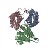


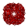
 Mass: 54.938 Da / Num. of mol.: 6 / Source method: obtained synthetically / Formula: Mn
 Mass: 207.124 Da / Num. of mol.: 3 / Source method: obtained synthetically / Formula: C5H10N3O4P
 Mass: 18.015 Da / Num. of mol.: 322 / Source method: isolated from a natural source / Formula: H2O
 Sample preparation
 / Beamline: I04 / Wavelength: 0.9795 Å
 Processing