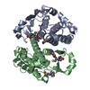
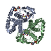
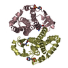
Entry Database : PDB / ID : 5djmTitle Structure of WT Human Glutathione Transferase in complex with cisplatin in the absence of glutathione. Glutathione S-transferase P Keywords / / Function / homology Function Domain/homology Component
/ / / / / / / / / / / / / / / / / / / / / / / / / / / / / / / / / / / / / / / / / / / / / / / / / / / / / / / / / / / / / / / / / / / / / / / / / / / / / / / / / / / / / / / / / / / Biological species Homo sapiens (human)Method / / / / Resolution : 1.9 Å Authors Parker, L.J. Journal : Proc.Natl.Acad.Sci.USA / Year : 2019Title : A structure-based mechanism of cisplatin resistance mediated by Glutathione Transferase P1-1Authors : Parker, L.J. / Italiano, L.C. / Hancock, N.C. / Aitken, J. / Harris, H.H. / Hansen, G. / Ascher, D.B. / Morton, C.J. / Parker, M.W. / Lo Bello, M. History Deposition Sep 2, 2015 Deposition site / Processing site Revision 1.0 Nov 2, 2016 Provider / Type Revision 1.1 Nov 22, 2017 Group / Refinement description / Category / softwareItem / _software.classificationRevision 1.2 Jun 19, 2019 Group / Database references / Category Item _citation.country / _citation.journal_abbrev ... _citation.country / _citation.journal_abbrev / _citation.journal_id_ASTM / _citation.journal_id_CSD / _citation.journal_id_ISSN / _citation.title / _citation.year Revision 1.3 Sep 27, 2023 Group Data collection / Database references ... Data collection / Database references / Derived calculations / Refinement description Category chem_comp_atom / chem_comp_bond ... chem_comp_atom / chem_comp_bond / database_2 / pdbx_initial_refinement_model / struct_conn Item _database_2.pdbx_DOI / _database_2.pdbx_database_accession ... _database_2.pdbx_DOI / _database_2.pdbx_database_accession / _struct_conn.ptnr1_auth_asym_id / _struct_conn.ptnr1_auth_comp_id / _struct_conn.ptnr1_auth_seq_id / _struct_conn.ptnr1_label_asym_id / _struct_conn.ptnr1_label_atom_id / _struct_conn.ptnr1_label_comp_id / _struct_conn.ptnr1_label_seq_id / _struct_conn.ptnr2_auth_asym_id / _struct_conn.ptnr2_auth_comp_id / _struct_conn.ptnr2_auth_seq_id / _struct_conn.ptnr2_label_asym_id / _struct_conn.ptnr2_label_atom_id / _struct_conn.ptnr2_label_comp_id / _struct_conn.ptnr2_label_seq_id
Show all Show less
 Movie
Movie Controller

 Yorodumi
 Open data
 Basic information
 Components
 Keywords
 Function and homology information
 S-nitrosoglutathione binding / nitric oxide storage / negative regulation of biosynthetic process / TRAF2-GSTP1 complex / kinase regulator activity / negative regulation of leukocyte proliferation / dinitrosyl-iron complex binding / common myeloid progenitor cell proliferation / hepoxilin biosynthetic process / glutathione derivative biosynthetic process ...
 Authors
 Citation
 Structure visualization
 Downloads & links
 Other downloads
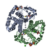


 PDBj
PDBj


 Assembly


 Mass: 195.237 Da / Num. of mol.: 2 / Source method: obtained synthetically / Formula: C6H13NO4S / Comment: pH buffer*YM
 Mass: 195.078 Da / Num. of mol.: 3 / Source method: obtained synthetically / Formula: Pt
 Mass: 18.015 Da / Num. of mol.: 105 / Source method: isolated from a natural source / Formula: H2O
 Sample preparation
 / Beamline: 14-BM-C / Wavelength: 0.9 Å
 Processing