 Movie
Movie Controller

[English] 日本語
 Yorodumi
- PDB-5dfy: Structure of the parental state of GAF3 from Slr1393 of Synechocy... -
+ Open data

- Basic information
| Entry | Database: PDB / ID: 5dfy | |||||||||
|---|---|---|---|---|---|---|---|---|---|---|
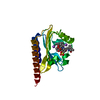
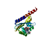
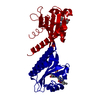
| Title | Structure of the parental state of GAF3 from Slr1393 of Synechocystis sp. PCC6803 (in vitro assembled protein/chromophore) | |||||||||
 Components Components | Histidine kinase | |||||||||
 Keywords Keywords | TRANSFERASE / cyanobacteriochrome / phycocyanobilin chromophore / photochromicity / bilin-binding GAF domain / photochrome-related protein | |||||||||
| Function / homology |  Function and homology information Function and homology information | |||||||||
| Biological species |  | |||||||||
| Method | X-RAY DIFFRACTION / SYNCHROTRON / MOLECULAR REPLACEMENT / Resolution: 1.6 Å | |||||||||
 Authors Authors | Xu, X.-L. / Zhao, K.-H. / Gaertner, W. / Hoeppner, A. | |||||||||
 Citation Citation | Journal: Proc.Natl.Acad.Sci.USA / Year: 2020 Title: Structural elements regulating the photochromicity in a cyanobacteriochrome Authors: Xu, X.-L. / Hoeppner, A. / Wiebeler, C. / Zhao, K.-H. / Schapiro, I. / Gaertner, W. | |||||||||
| History |
|
- Structure visualization
| Structure viewer | Molecule: MolmilJmol/JSmol |
|---|
- Downloads & links
-Download
| PDBx/mmCIF format | 5dfy.cif.gz | 51.7 KB | Display | PDBx/mmCIF format |
|---|---|---|---|---|
| PDB format | pdb5dfy.ent.gz | 38 KB | Display | PDB format |
| PDBx/mmJSON format | 5dfy.json.gz | Tree view | PDBx/mmJSON format | |
| Others |  Other downloads Other downloads |
-Validation report
| Arichive directory | https://data.pdbj.org/pub/pdb/validation_reports/df/5dfyftp://data.pdbj.org/pub/pdb/validation_reports/df/5dfy | HTTPS FTP |
|---|
-Related structure data


-Links
 PDBj
PDBj


- Assembly
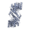
| Deposited unit | 
| ||||||||
|---|---|---|---|---|---|---|---|---|---|
| 1 | 
| ||||||||
| Unit cell |
| ||||||||
| Components on special symmetry positions |
|
-Components
| #1: Protein | Mass: 24856.637 Da / Num. of mol.: 1 / Fragment: residues 441-597 Source method: isolated from a genetically manipulated source Source: (gene. exp.) Gene: slr1393 / Production host: Escherichia coli BL21(DE3) (bacteria) / References: UniProt: P73184, histidine kinase |
|---|---|
| #2: Chemical | ChemComp-CYC / Phycocyanobilin  Mass: 588.694 Da / Num. of mol.: 1 / Source method: obtained synthetically / Formula: C33H40N4O6 Mass: 588.694 Da / Num. of mol.: 1 / Source method: obtained synthetically / Formula: C33H40N4O6 |
| #3: Water | ChemComp-HOH / Water Mass: 18.015 Da / Num. of mol.: 219 / Source method: isolated from a natural source / Formula: H2O Mass: 18.015 Da / Num. of mol.: 219 / Source method: isolated from a natural source / Formula: H2O |
-Experimental details
-Experiment
| Experiment | Method: X-RAY DIFFRACTION |
|---|
- Sample preparation
| Crystal | Density Matthews: 2.38 Å3/Da / Density % sol: 48.27 % |
|---|---|
| Crystal grow | Temperature: 285 K / Method: vapor diffusion, hanging drop / pH: 5.6 Details: 0.02 M sodium citrate pH 5.6, 0.1 M sodium chloride, 11 % (w/v) PEG 3350 |
-Data collection
| Diffraction | Mean temperature: 100 K |
|---|---|
| Diffraction source | Source: SYNCHROTRON / Site: ESRF  / Beamline: ID23-1 / Wavelength: 1.00894 Å / Beamline: ID23-1 / Wavelength: 1.00894 Å |
| Detector | Type: DECTRIS PILATUS 6M / Detector: PIXEL / Date: Mar 2, 2015 |
| Radiation | Protocol: SINGLE WAVELENGTH / Monochromatic (M) / Laue (L): M / Scattering type: x-ray |
| Radiation wavelength | Wavelength: 1.00894 Å / Relative weight: 1 |
| Reflection | Resolution: 1.6→44.51 Å / Num. obs: 32428 / % possible obs: 99.9 % / Redundancy: 12.71 % / Rsym value: 0.05 / Net I/σ(I): 27.87 |
| Reflection shell | Resolution: 1.6→1.7 Å / Redundancy: 12.4 % / Mean I/σ(I) obs: 6.44 / Rsym value: 0.342 / % possible all: 99.5 |
- Processing
| Software |
| |||||||||||||||||||||||||||||||||||||||||||||||||||||||||||||||||||||||||||||||||||||||||||
|---|---|---|---|---|---|---|---|---|---|---|---|---|---|---|---|---|---|---|---|---|---|---|---|---|---|---|---|---|---|---|---|---|---|---|---|---|---|---|---|---|---|---|---|---|---|---|---|---|---|---|---|---|---|---|---|---|---|---|---|---|---|---|---|---|---|---|---|---|---|---|---|---|---|---|---|---|---|---|---|---|---|---|---|---|---|---|---|---|---|---|---|---|
| Refinement | Method to determine structure: MOLECULAR REPLACEMENT / Resolution: 1.6→33.629 Å / SU ML: 0.12 / Cross valid method: FREE R-VALUE / σ(F): 1.16 / Phase error: 16.51 / Stereochemistry target values: ML
| |||||||||||||||||||||||||||||||||||||||||||||||||||||||||||||||||||||||||||||||||||||||||||
| Solvent computation | Shrinkage radii: 0.9 Å / VDW probe radii: 1.11 Å / Solvent model: FLAT BULK SOLVENT MODEL | |||||||||||||||||||||||||||||||||||||||||||||||||||||||||||||||||||||||||||||||||||||||||||
| Refinement step | Cycle: LAST / Resolution: 1.6→33.629 Å
| |||||||||||||||||||||||||||||||||||||||||||||||||||||||||||||||||||||||||||||||||||||||||||
| Refine LS restraints |
| |||||||||||||||||||||||||||||||||||||||||||||||||||||||||||||||||||||||||||||||||||||||||||
| LS refinement shell |
|
