 Movie
Movie Controller

[English] 日本語
 Yorodumi
- PDB-5d67: Crystal structure of an EF-Hand calcium binding domain of CAP-Bin... -
+ Open data

- Basic information
| Entry | Database: PDB / ID: 5d67 | ||||||
|---|---|---|---|---|---|---|---|
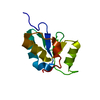
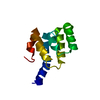
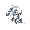
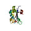
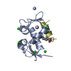
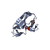
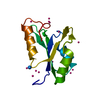
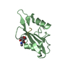
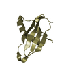
| Title | Crystal structure of an EF-Hand calcium binding domain of CAP-Binding Protein Complex-Interacting Protein 1 (EFCAB6) from Homo sapiens at 2.00 A resolution | ||||||
 Components Components | EF-hand calcium-binding domain-containing protein 6 | ||||||
 Keywords Keywords | METAL BINDING PROTEIN /  Structural Genomics / Joint Center for Structural Genomics / JCSG / Protein Structure Initiative / PSI-BIOLOGY / Partnership for Nuclear Receptor Signaling Code Biology / NHRS Structural Genomics / Joint Center for Structural Genomics / JCSG / Protein Structure Initiative / PSI-BIOLOGY / Partnership for Nuclear Receptor Signaling Code Biology / NHRS | ||||||
| Function / homology |  Function and homology information Function and homology informationcell projection / cytoskeleton / calcium ion binding / nucleoplasm / cytoplasmSimilarity search - Function | ||||||
| Biological species |  Homo sapiens (human) Homo sapiens (human) | ||||||
| Method | X-RAY DIFFRACTION / SYNCHROTRON / SAD / Resolution: 2 Å | ||||||
 Authors Authors | Joint Center for Structural Genomics (JCSG) / Partnership for Nuclear Receptor Signaling Code Biology / Partnership for Nuclear Receptor Signaling Code Biology (NHRS) | ||||||
 Citation Citation | Journal: To be published Title: Crystal structure of an EF-Hand calcium binding domain of CAP-Binding Protein Complex-Interacting Protein 1 (EFCAB6) from Homo sapiens at 2.00 A resolution Authors: Joint Center for Structural Genomics (JCSG) / Partnership for Nuclear Receptor Signaling Code Biology | ||||||
| History |
|
- Structure visualization
| Structure viewer | Molecule: MolmilJmol/JSmol |
|---|
- Downloads & links
-Download
| PDBx/mmCIF format | 5d67.cif.gz | 164.7 KB | Display | PDBx/mmCIF format |
|---|---|---|---|---|
| PDB format | pdb5d67.ent.gz | 133.4 KB | Display | PDB format |
| PDBx/mmJSON format | 5d67.json.gz | Tree view | PDBx/mmJSON format | |
| Others |  Other downloads Other downloads |
-Validation report
| Arichive directory | https://data.pdbj.org/pub/pdb/validation_reports/d6/5d67ftp://data.pdbj.org/pub/pdb/validation_reports/d6/5d67 | HTTPS FTP |
|---|
-Related structure data
| Similar structure data | |
|---|---|
| Other databases |

-Links
 PDBj
PDBj

- Assembly
| Deposited unit | 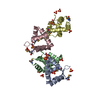
| ||||||||
|---|---|---|---|---|---|---|---|---|---|
| 1 | 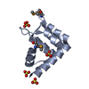
| ||||||||
| 2 | 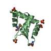
| ||||||||
| 3 | 
| ||||||||
| 4 | 
| ||||||||
| Unit cell |
|
-Components
| #1: Protein | Mass: 11778.729 Da / Num. of mol.: 4 / Fragment: UNP residues 1160-1260 / Mutation: D1218G Source method: isolated from a genetically manipulated source Source: (gene. exp.) Homo sapiens (human) / Gene: EFCAB6, DJBP, KIAA1672 / Plasmid: SpeedET / Production host:  Escherichia Coli (E. coli) / Strain (production host): PB1 / References: UniProt: Q5THR3 Escherichia Coli (E. coli) / Strain (production host): PB1 / References: UniProt: Q5THR3#2: Chemical | ChemComp-SO4 / Sulfate  Mass: 96.063 Da / Num. of mol.: 15 / Source method: obtained synthetically / Formula: SO4 Mass: 96.063 Da / Num. of mol.: 15 / Source method: obtained synthetically / Formula: SO4#3: Water | ChemComp-HOH / | Water Mass: 18.015 Da / Num. of mol.: 328 / Source method: isolated from a natural source / Formula: H2O Mass: 18.015 Da / Num. of mol.: 328 / Source method: isolated from a natural source / Formula: H2O |
|---|
-Experimental details
-Experiment
| Experiment | Method: X-RAY DIFFRACTION / Number of used crystals: 1 |
|---|
- Sample preparation
| Crystal | Density Matthews: 2.15 Å3/Da / Density % sol: 42.91 % |
|---|---|
| Crystal grow | Temperature: 277 K / Method: vapor diffusion, sitting drop Details: 30% polyethylene glycol 4000, 0.2M ammonium sulfate |
-Data collection
| Diffraction | Mean temperature: 100 K | |||||||||||||||||||||||||||||||||||||||||||||||||||||||||||||||||||||||||||||||||||||||||||||||||||||||||||||||||||||||||
|---|---|---|---|---|---|---|---|---|---|---|---|---|---|---|---|---|---|---|---|---|---|---|---|---|---|---|---|---|---|---|---|---|---|---|---|---|---|---|---|---|---|---|---|---|---|---|---|---|---|---|---|---|---|---|---|---|---|---|---|---|---|---|---|---|---|---|---|---|---|---|---|---|---|---|---|---|---|---|---|---|---|---|---|---|---|---|---|---|---|---|---|---|---|---|---|---|---|---|---|---|---|---|---|---|---|---|---|---|---|---|---|---|---|---|---|---|---|---|---|---|---|---|
| Diffraction source | Source: SYNCHROTRON / Site: SSRL  / Beamline: BL14-1 / Wavelength: 0.97917 Å / Beamline: BL14-1 / Wavelength: 0.97917 Å | |||||||||||||||||||||||||||||||||||||||||||||||||||||||||||||||||||||||||||||||||||||||||||||||||||||||||||||||||||||||||
| Detector | Type: MARMOSAIC 325 mm CCD / Detector: CCD / Date: Mar 8, 2015 Details: Vertical focusing mirror; double crystal Si(111) monochromator | |||||||||||||||||||||||||||||||||||||||||||||||||||||||||||||||||||||||||||||||||||||||||||||||||||||||||||||||||||||||||
| Radiation | Monochromator: double crystal Si(111) / Protocol: SINGLE WAVELENGTH / Monochromatic (M) / Laue (L): M / Scattering type: x-ray | |||||||||||||||||||||||||||||||||||||||||||||||||||||||||||||||||||||||||||||||||||||||||||||||||||||||||||||||||||||||||
| Radiation wavelength | Wavelength: 0.97917 Å / Relative weight: 1 | |||||||||||||||||||||||||||||||||||||||||||||||||||||||||||||||||||||||||||||||||||||||||||||||||||||||||||||||||||||||||
| Reflection | Resolution: 2→27.118 Å / Num. obs: 26723 / % possible obs: 98.1 % / Observed criterion σ(I): -3 / Redundancy: 3.787 % / Biso Wilson estimate: 26.457 Å2 / Rmerge F obs: 0.996 / Rmerge(I) obs: 0.112 / Rrim(I) all: 0.13 / Net I/σ(I): 10.59 / Num. measured all: 197737 | |||||||||||||||||||||||||||||||||||||||||||||||||||||||||||||||||||||||||||||||||||||||||||||||||||||||||||||||||||||||||
| Reflection shell |
|
-Phasing
| Phasing | Method: SAD |
|---|
- Processing
| Software |
| |||||||||||||||||||||||||||||||||||||||||||||||||||||||||||||||||||||||||||||||||||||||||||||||||||||||||||||||||||||||||||||
|---|---|---|---|---|---|---|---|---|---|---|---|---|---|---|---|---|---|---|---|---|---|---|---|---|---|---|---|---|---|---|---|---|---|---|---|---|---|---|---|---|---|---|---|---|---|---|---|---|---|---|---|---|---|---|---|---|---|---|---|---|---|---|---|---|---|---|---|---|---|---|---|---|---|---|---|---|---|---|---|---|---|---|---|---|---|---|---|---|---|---|---|---|---|---|---|---|---|---|---|---|---|---|---|---|---|---|---|---|---|---|---|---|---|---|---|---|---|---|---|---|---|---|---|---|---|---|
| Refinement | Method to determine structure: SAD / Resolution: 2→27.118 Å / Cor.coef. Fo:Fc: 0.953 / Cor.coef. Fo:Fc free: 0.9373 / Occupancy max: 1 / Occupancy min: 0.37 / Cross valid method: THROUGHOUT / σ(F): 0 Details: 1. ATOM RECORDS CONTAIN SUM OF TLS AND RESIDUAL B FACTORS. 2. ANISOU RECORDS CONTAIN SUM OF TLS AND RESIDUAL U FACTORS. 3. A MET-INHIBITION PROTOCOL WAS USED FOR SELENOMETHIONINE ...Details: 1. ATOM RECORDS CONTAIN SUM OF TLS AND RESIDUAL B FACTORS. 2. ANISOU RECORDS CONTAIN SUM OF TLS AND RESIDUAL U FACTORS. 3. A MET-INHIBITION PROTOCOL WAS USED FOR SELENOMETHIONINE INCORPORATION DURING PROTEIN EXPRESSION. THE OCCUPANCY OF THE SE ATOMS IN THE MSE RESIDUES WAS REDUCED TO 0.75 FOR THE REDUCED SCATTERING POWER DUE TO PARTIAL S-MET INCORPORATION. 4. THE SAD PHASES WERE USED AS RESTRAINTS DURING REFINEMENT
| |||||||||||||||||||||||||||||||||||||||||||||||||||||||||||||||||||||||||||||||||||||||||||||||||||||||||||||||||||||||||||||
| Displacement parameters | Biso max: 161.22 Å2 / Biso mean: 35.3461 Å2 / Biso min: 10.12 Å2
| |||||||||||||||||||||||||||||||||||||||||||||||||||||||||||||||||||||||||||||||||||||||||||||||||||||||||||||||||||||||||||||
| Refine analyze | Luzzati coordinate error obs: 0.209 Å | |||||||||||||||||||||||||||||||||||||||||||||||||||||||||||||||||||||||||||||||||||||||||||||||||||||||||||||||||||||||||||||
| Refinement step | Cycle: LAST / Resolution: 2→27.118 Å
| |||||||||||||||||||||||||||||||||||||||||||||||||||||||||||||||||||||||||||||||||||||||||||||||||||||||||||||||||||||||||||||
| Refine LS restraints |
| |||||||||||||||||||||||||||||||||||||||||||||||||||||||||||||||||||||||||||||||||||||||||||||||||||||||||||||||||||||||||||||
| LS refinement shell | Resolution: 2→2.08 Å / Total num. of bins used: 13
| |||||||||||||||||||||||||||||||||||||||||||||||||||||||||||||||||||||||||||||||||||||||||||||||||||||||||||||||||||||||||||||
| Refinement TLS params. | Method: refined / Refine-ID: X-RAY DIFFRACTION
| |||||||||||||||||||||||||||||||||||||||||||||||||||||||||||||||||||||||||||||||||||||||||||||||||||||||||||||||||||||||||||||
| Refinement TLS group |
|
