- PDB-5d1y: Low resolution crystal structure of human ribonucleotide reductas... -
+
Open data
ID or keywords:
Loading...
-
Basic information
Entry
Database: PDB / ID: 5d1y
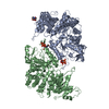
Title
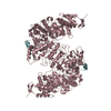
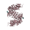
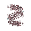
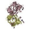
Low resolution crystal structure of human ribonucleotide reductase alpha6 hexamer in complex with dATP
Components
Ribonucleoside-diphosphate reductase large subunitRibonucleotide reductase
Keywords
OXIDOREDUCTASE / deoxyribonucleotide / oligomerization / ATP cone
Function / homology
Function and homology information
ribonucleoside-diphosphate reductase activity / pyrimidine nucleobase metabolic process / cell proliferation in forebrain / positive regulation of G0 to G1 transition / mitochondrial DNA replication / ribonucleoside diphosphate metabolic process / 2'-deoxyribonucleotide biosynthetic process / ribonucleoside-diphosphate reductase complex / ribonucleoside-diphosphate reductase / ribonucleoside-diphosphate reductase activity, thioredoxin disulfide as acceptor ...ribonucleoside-diphosphate reductase activity / pyrimidine nucleobase metabolic process / cell proliferation in forebrain / positive regulation of G0 to G1 transition / mitochondrial DNA replication / ribonucleoside diphosphate metabolic process / 2'-deoxyribonucleotide biosynthetic process / ribonucleoside-diphosphate reductase complex / ribonucleoside-diphosphate reductase / ribonucleoside-diphosphate reductase activity, thioredoxin disulfide as acceptor / Interconversion of nucleotide di- and triphosphates / deoxyribonucleotide biosynthetic process / protein heterotetramerization / response to ionizing radiation / DNA synthesis involved in DNA repair / positive regulation of G1/S transition of mitotic cell cycle / positive regulation of G2/M transition of mitotic cell cycle / cell projection / male gonad development / disordered domain specific binding / retina development in camera-type eye / nuclear envelope / DNA repair / neuronal cell body / mitochondrion / ATP binding / identical protein binding / cytosol Similarity search - Function
Ribonucleotide reductase, class I , alpha subunit / Ribonucleotide reductase large subunit signature. / Ribonucleoside-diphosphate reductase large subunit / ATP-cone domain / ATP cone domain / ATP-cone domain profile. / Ribonucleotide reductase R1 subunit, N-terminal / Ribonucleotide reductase large subunit, N-terminal / Ribonucleotide reductase, all-alpha domain / Ribonucleotide reductase large subunit, C-terminal / Ribonucleotide reductase, barrel domain Similarity search - Domain/homology
This structure has undergone only rigid body and group B-factor refinement. The data-to-parameter ...This structure has undergone only rigid body and group B-factor refinement. The data-to-parameter ratio (3405 reflections, 11559 atoms) was not sufficient for individual atom position or B-factor refinement.
In the structure databanks used in Yorodumi, some data are registered as the other names, "COVID-19 virus" and "2019-nCoV". Here are the details of the virus and the list of structure data.
Jan 31, 2019. EMDB accession codes are about to change! (news from PDBe EMDB page)
EMDB accession codes are about to change! (news from PDBe EMDB page)
The allocation of 4 digits for EMDB accession codes will soon come to an end. Whilst these codes will remain in use, new EMDB accession codes will include an additional digit and will expand incrementally as the available range of codes is exhausted. The current 4-digit format prefixed with “EMD-” (i.e. EMD-XXXX) will advance to a 5-digit format (i.e. EMD-XXXXX), and so on. It is currently estimated that the 4-digit codes will be depleted around Spring 2019, at which point the 5-digit format will come into force.
The EM Navigator/Yorodumi systems omit the EMD- prefix.
Related info.:Q: What is EMD? / ID/Accession-code notation in Yorodumi/EM Navigator
Yorodumi is a browser for structure data from EMDB, PDB, SASBDB, etc.
This page is also the successor to EM Navigator detail page, and also detail information page/front-end page for Omokage search.
The word "yorodu" (or yorozu) is an old Japanese word meaning "ten thousand". "mi" (miru) is to see.
Related info.:EMDB / PDB / SASBDB / Comparison of 3 databanks / Yorodumi Search / Aug 31, 2016. New EM Navigator & Yorodumi / Yorodumi Papers / Jmol/JSmol / Function and homology information / Changes in new EM Navigator and Yorodumi
 Movie
Movie Controller

 Yorodumi
 Open data
 Basic information
 Components
 Ribonucleotide reductase
  Keywords
 Function and homology information
 Authors
 United States, 5items
  Citation
 Structure visualization
 Downloads & links
 Other downloads
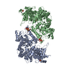


 PDBj
PDBj

 Assembly
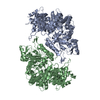

 Sample preparation
 Processing