 Movie
Movie Controller

[English] 日本語
 Yorodumi
- PDB-5cm8: Structural Basis for the Selectivity of Guanine Nucleotide Exchan... -
+ Open data

- Basic information
| Entry | Database: PDB / ID: 5cm8 | ||||||
|---|---|---|---|---|---|---|---|
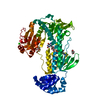
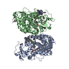
| Title | Structural Basis for the Selectivity of Guanine Nucleotide Exchange Factors for the small G-protein Ral | ||||||
 Components Components |
| ||||||
 Keywords Keywords |  SIGNALING PROTEIN / Complex G-protein Exchange Factor SIGNALING PROTEIN / Complex G-protein Exchange Factor | ||||||
| Function / homology |  Function and homology information Function and homology informationR3/R4 cell fate commitment / regulation of Ral protein signal transduction / border follicle cell migration / negative regulation of JNK cascade / regulation of cell morphogenesis / Flemming body / negative regulation of cardiac muscle cell apoptotic process / positive regulation of epidermal growth factor receptor signaling pathway / small GTPase-mediated signal transduction / positive regulation of stem cell proliferation ...R3/R4 cell fate commitment / regulation of Ral protein signal transduction / border follicle cell migration / negative regulation of JNK cascade / regulation of cell morphogenesis / Flemming body / negative regulation of cardiac muscle cell apoptotic process / positive regulation of epidermal growth factor receptor signaling pathway / small GTPase-mediated signal transduction / positive regulation of stem cell proliferation / cleavage furrow / negative regulation of innate immune response / guanyl-nucleotide exchange factor activity / small monomeric GTPase / G protein activity / PDZ domain binding / receptor internalization / GDP binding / Ras protein signal transduction / defense response to Gram-negative bacterium / positive regulation of phosphatidylinositol 3-kinase/protein kinase B signal transduction / innate immune response / GTPase activity / GTP binding / plasma membraneSimilarity search - Function | ||||||
| Biological species |  Mus musculus (house mouse) Mus musculus (house mouse) Drosophila melanogaster (fruit fly) Drosophila melanogaster (fruit fly) | ||||||
| Method | X-RAY DIFFRACTION / SYNCHROTRON / MOLECULAR REPLACEMENT / Resolution: 2.6 Å | ||||||
 Authors Authors | Popovic, M. / Schouten, A. / Rehmann, H. | ||||||
 Citation Citation | Journal: J.Struct.Biol. / Year: 2016 Title: The structure of the Guanine Nucleotide Exchange Factor Rlf in complex with the small G-protein Ral identifies conformational intermediates of the exchange reaction and the basis for the selectivity. Authors: Popovic, M. / Schouten, A. / Rensen-de Leeuw, M. / Rehmann, H. | ||||||
| History |
|
- Structure visualization
| Structure viewer | Molecule: MolmilJmol/JSmol |
|---|
- Downloads & links
-Download
| PDBx/mmCIF format | 5cm8.cif.gz | 129 KB | Display | PDBx/mmCIF format |
|---|---|---|---|---|
| PDB format | pdb5cm8.ent.gz | 97.2 KB | Display | PDB format |
| PDBx/mmJSON format | 5cm8.json.gz | Tree view | PDBx/mmJSON format | |
| Others |  Other downloads Other downloads |
-Validation report
| Arichive directory | https://data.pdbj.org/pub/pdb/validation_reports/cm/5cm8ftp://data.pdbj.org/pub/pdb/validation_reports/cm/5cm8 | HTTPS FTP |
|---|
-Related structure data
| Related structure data |  5cm9C  4jgwS C: citing same article ( S: Starting model for refinement |
|---|---|
| Similar structure data |

-Links
 PDBj
PDBj


- Assembly
| Deposited unit | 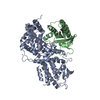
| ||||||||
|---|---|---|---|---|---|---|---|---|---|
| 1 |
| ||||||||
| Unit cell |
|
-Components
| #1: Protein | Mass: 52113.898 Da / Num. of mol.: 1 / Fragment: UNP residues 50-514 Source method: isolated from a genetically manipulated source Source: (gene. exp.) Mus musculus (house mouse) / Gene: Rgl2, Rab2l, Rlf / Plasmid: pGEX6P3 / Production host:  Escherichia coli (E. coli) / Strain (production host): CK600 / References: UniProt: Q61193 Escherichia coli (E. coli) / Strain (production host): CK600 / References: UniProt: Q61193 |
|---|---|
| #2: Protein | Mass: 23117.285 Da / Num. of mol.: 1 Source method: isolated from a genetically manipulated source Source: (gene. exp.) Drosophila melanogaster (fruit fly) / Gene: Rala, CG2849 / Plasmid: pGEX4T3 / Production host: Escherichia coli (E. coli) / Strain (production host): CK600 / References: UniProt: P48555 |
| #3: Water | ChemComp-HOH / Water Mass: 18.015 Da / Num. of mol.: 45 / Source method: isolated from a natural source / Formula: H2O Mass: 18.015 Da / Num. of mol.: 45 / Source method: isolated from a natural source / Formula: H2O |
-Experimental details
-Experiment
| Experiment | Method: X-RAY DIFFRACTION |
|---|
- Sample preparation
| Crystal | Density Matthews: 2.59 Å3/Da / Density % sol: 52.56 % |
|---|---|
| Crystal grow | Temperature: 289 K / Method: vapor diffusion, sitting drop / pH: 7.45 Details: 15% PEG 3350, 200 mM ammonium acetate, 100 mM Bis-Tris propane |
-Data collection
| Diffraction | Mean temperature: 100 K |
|---|---|
| Diffraction source | Source: SYNCHROTRON / Site: ESRF  / Beamline: ID23-1 / Wavelength: 0.95372 Å / Beamline: ID23-1 / Wavelength: 0.95372 Å |
| Detector | Type: ADSC QUANTUM 315r / Detector: CCD / Date: Nov 25, 2012 |
| Radiation | Protocol: SINGLE WAVELENGTH / Monochromatic (M) / Laue (L): M / Scattering type: x-ray |
| Radiation wavelength | Wavelength: 0.95372 Å / Relative weight: 1 |
| Reflection | Resolution: 2.6→50 Å / Num. all: 71227 / Num. obs: 23162 / % possible obs: 97.7 % / Redundancy: 3.1 % / Rmerge(I) obs: 0.096 / Net I/σ(I): 15.81 |
| Reflection shell | Resolution: 2.6→2.7 Å / Redundancy: 3.2 % / Mean I/σ(I) obs: 3.21 / % possible all: 99.6 |
- Processing
| Software |
| ||||||||||||||||||||||||||||||||||||||||||||||||||||||||||||||||||||||||||||||||||||||||||||||||||||||||||||||||||||||||||||||||||||||||||||||||||||||||||||||||||||||||||||||||||||||
|---|---|---|---|---|---|---|---|---|---|---|---|---|---|---|---|---|---|---|---|---|---|---|---|---|---|---|---|---|---|---|---|---|---|---|---|---|---|---|---|---|---|---|---|---|---|---|---|---|---|---|---|---|---|---|---|---|---|---|---|---|---|---|---|---|---|---|---|---|---|---|---|---|---|---|---|---|---|---|---|---|---|---|---|---|---|---|---|---|---|---|---|---|---|---|---|---|---|---|---|---|---|---|---|---|---|---|---|---|---|---|---|---|---|---|---|---|---|---|---|---|---|---|---|---|---|---|---|---|---|---|---|---|---|---|---|---|---|---|---|---|---|---|---|---|---|---|---|---|---|---|---|---|---|---|---|---|---|---|---|---|---|---|---|---|---|---|---|---|---|---|---|---|---|---|---|---|---|---|---|---|---|---|---|
| Refinement | Method to determine structure: MOLECULAR REPLACEMENT Starting model: 4JGW as poly Ala model Resolution: 2.6→44.42 Å / Cor.coef. Fo:Fc: 0.919 / Cor.coef. Fo:Fc free: 0.864 / SU B: 15.198 / SU ML: 0.329 / Cross valid method: THROUGHOUT / ESU R: 0.792 / ESU R Free: 0.391 / Stereochemistry target values: MAXIMUM LIKELIHOOD / Details: HYDROGENS HAVE BEEN ADDED IN THE RIDING POSITIONS
| ||||||||||||||||||||||||||||||||||||||||||||||||||||||||||||||||||||||||||||||||||||||||||||||||||||||||||||||||||||||||||||||||||||||||||||||||||||||||||||||||||||||||||||||||||||||
| Solvent computation | Ion probe radii: 0.8 Å / Shrinkage radii: 0.8 Å / VDW probe radii: 1.4 Å / Solvent model: MASK | ||||||||||||||||||||||||||||||||||||||||||||||||||||||||||||||||||||||||||||||||||||||||||||||||||||||||||||||||||||||||||||||||||||||||||||||||||||||||||||||||||||||||||||||||||||||
| Displacement parameters | Biso mean: 64.994 Å2
| ||||||||||||||||||||||||||||||||||||||||||||||||||||||||||||||||||||||||||||||||||||||||||||||||||||||||||||||||||||||||||||||||||||||||||||||||||||||||||||||||||||||||||||||||||||||
| Refinement step | Cycle: 1 / Resolution: 2.6→44.42 Å
| ||||||||||||||||||||||||||||||||||||||||||||||||||||||||||||||||||||||||||||||||||||||||||||||||||||||||||||||||||||||||||||||||||||||||||||||||||||||||||||||||||||||||||||||||||||||
| Refine LS restraints |
|