 Movie
Movie Controller

[English] 日本語
 Yorodumi
- PDB-1k2o: Cytochrome P450Cam with Bound BIS(2,2'-BIPYRIDINE)-(5-METHYL-2-2'... -
+ Open data

- Basic information
| Entry | Database: PDB / ID: 1k2o | ||||||
|---|---|---|---|---|---|---|---|
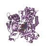
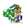
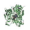
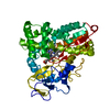
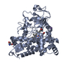
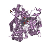
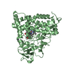
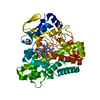
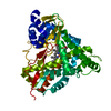
| Title | Cytochrome P450Cam with Bound BIS(2,2'-BIPYRIDINE)-(5-METHYL-2-2'-BIPYRIDINE)-C2-ADAMANTANE RUTHENIUM (II) | ||||||
 Components Components | Cytochrome P450CAM | ||||||
 Keywords Keywords |  OXIDOREDUCTASE / P450 / monooxygenase / electron transfer / energy transfer / fluorinated aromatics / biphenyl / adamantane / ruthenium channel / substrate-binding OXIDOREDUCTASE / P450 / monooxygenase / electron transfer / energy transfer / fluorinated aromatics / biphenyl / adamantane / ruthenium channel / substrate-binding | ||||||
| Function / homology |  Function and homology informationcamphor 5-monooxygenase / camphor 5-monooxygenase activity / (+)-camphor catabolic process / cholest-4-en-3-one 26-monooxygenase activity / steroid hydroxylase activity / cholesterol catabolic process / iron ion binding / heme binding / cytoplasm Function and homology informationcamphor 5-monooxygenase / camphor 5-monooxygenase activity / (+)-camphor catabolic process / cholest-4-en-3-one 26-monooxygenase activity / steroid hydroxylase activity / cholesterol catabolic process / iron ion binding / heme binding / cytoplasmSimilarity search - Function | ||||||
| Biological species |  Pseudomonas putida (bacteria) Pseudomonas putida (bacteria) | ||||||
| Method | X-RAY DIFFRACTION / SYNCHROTRON / MOLECULAR REPLACEMENT / Resolution: 1.65 Å | ||||||
 Authors Authors | Dunn, A.R. / Dmochowski, I.J. / Bilwes, A.M. / Gray, H.B. / Crane, B.R. | ||||||
 Citation Citation | Journal: Proc.Natl.Acad.Sci.USA / Year: 2001 Title: Probing the open state of cytochrome P450cam with ruthenium-linker substrates. Authors: Dunn, A.R. / Dmochowski, I.J. / Bilwes, A.M. / Gray, H.B. / Crane, B.R. | ||||||
| History |
|
- Structure visualization
| Structure viewer | Molecule: MolmilJmol/JSmol |
|---|
- Downloads & links
-Download
| PDBx/mmCIF format | 1k2o.cif.gz | 202.2 KB | Display | PDBx/mmCIF format |
|---|---|---|---|---|
| PDB format | pdb1k2o.ent.gz | 158 KB | Display | PDB format |
| PDBx/mmJSON format | 1k2o.json.gz | Tree view | PDBx/mmJSON format | |
| Others |  Other downloads Other downloads |
-Validation report
| Arichive directory | https://data.pdbj.org/pub/pdb/validation_reports/k2/1k2oftp://data.pdbj.org/pub/pdb/validation_reports/k2/1k2o | HTTPS FTP |
|---|
-Related structure data
| Related structure data | 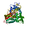 2cppS S: Starting model for refinement |
|---|---|
| Similar structure data |


-Links
 PDBj
PDBj


- Assembly
| Deposited unit | 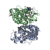
| ||||||||
|---|---|---|---|---|---|---|---|---|---|
| 1 | 
| ||||||||
| 2 | 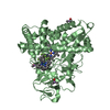
| ||||||||
| Unit cell |
|
-Components
-Protein , 1 types, 2 molecules AB
| #1: Protein | Mass: 46556.816 Da / Num. of mol.: 2 / Mutation: C334A Source method: isolated from a genetically manipulated source Source: (gene. exp.) Pseudomonas putida (bacteria) / Production host: Escherichia coli (E. coli) / Strain (production host): TBY / References: UniProt: P00183, camphor 5-monooxygenase |
|---|
-Non-polymers , 5 types, 705 molecules 


| #2: Chemical | ChemComp-CAC / Cacodylic acid Mass: 136.989 Da / Num. of mol.: 5 / Source method: obtained synthetically / Formula: C2H6AsO2 Mass: 136.989 Da / Num. of mol.: 5 / Source method: obtained synthetically / Formula: C2H6AsO2#3: Chemical | Heme B Mass: 616.487 Da / Num. of mol.: 2 / Source method: obtained synthetically / Formula: C34H32FeN4O4 Mass: 616.487 Da / Num. of mol.: 2 / Source method: obtained synthetically / Formula: C34H32FeN4O4#4: Chemical |  Mass: 1075.277 Da / Num. of mol.: 2 / Source method: obtained synthetically / Formula: C54H75F8N7Ru Mass: 1075.277 Da / Num. of mol.: 2 / Source method: obtained synthetically / Formula: C54H75F8N7Ru#5: Chemical |  Mass: 1075.277 Da / Num. of mol.: 2 / Source method: obtained synthetically / Formula: C54H75F8N7Ru Mass: 1075.277 Da / Num. of mol.: 2 / Source method: obtained synthetically / Formula: C54H75F8N7Ru#6: Water | ChemComp-HOH / | WaterMass: 18.015 Da / Num. of mol.: 694 / Source method: isolated from a natural source / Formula: H2O |
|---|
-Experimental details
-Experiment
| Experiment | Method: X-RAY DIFFRACTION / Number of used crystals: 1 |
|---|
- Sample preparation
| Crystal | Density Matthews: 2.65 Å3/Da / Density % sol: 53.5 % | ||||||||||||||||||||||||||||||||||||||||||||||||
|---|---|---|---|---|---|---|---|---|---|---|---|---|---|---|---|---|---|---|---|---|---|---|---|---|---|---|---|---|---|---|---|---|---|---|---|---|---|---|---|---|---|---|---|---|---|---|---|---|---|
| Crystal grow | Temperature: 277 K / Method: slow cooling / pH: 7.5 Details: Hepes, PEG, KCL, DTT, pH 7.5, slow cooling, temperature 277K | ||||||||||||||||||||||||||||||||||||||||||||||||
| Crystal grow | *PLUS Temperature: 4 ℃ / Method: vapor diffusion, hanging drop | ||||||||||||||||||||||||||||||||||||||||||||||||
| Components of the solutions | *PLUS
|
-Data collection
| Diffraction | Mean temperature: 100 K |
|---|---|
| Diffraction source | Source: SYNCHROTRON / Site: SSRL  / Beamline: BL9-1 / Wavelength: 0.72 Å / Beamline: BL9-1 / Wavelength: 0.72 Å |
| Detector | Type: MARRESEARCH / Detector: IMAGE PLATE / Date: May 24, 2000 |
| Radiation | Monochromator: Si / Protocol: SINGLE WAVELENGTH / Monochromatic (M) / Laue (L): M / Scattering type: x-ray |
| Radiation wavelength | Wavelength: 0.72 Å / Relative weight: 1 |
| Reflection | Resolution: 1.65→28 Å / Num. all: 111741 / Num. obs: 111741 / % possible obs: 97.8 % / Observed criterion σ(F): 0 / Observed criterion σ(I): 0 / Biso Wilson estimate: 19 Å2 / Rsym value: 0.038 / Net I/σ(I): 16.4 |
| Reflection shell | Resolution: 1.65→1.71 Å / Mean I/σ(I) obs: 2 / Rsym value: 0.292 / % possible all: 97 |
| Reflection | *PLUS Rmerge(I) obs: 0.038 |
| Reflection shell | *PLUS % possible obs: 97 % / Rmerge(I) obs: 0.292 |
- Processing
| Software |
| ||||||||||||||||||||||||||||||||||||||||
|---|---|---|---|---|---|---|---|---|---|---|---|---|---|---|---|---|---|---|---|---|---|---|---|---|---|---|---|---|---|---|---|---|---|---|---|---|---|---|---|---|---|
| Refinement | Method to determine structure: MOLECULAR REPLACEMENT Starting model: 2cpp Resolution: 1.65→19.81 Å / Rfactor Rfree error: 0.002 / Data cutoff high absF: 819940.02 / Data cutoff low absF: 0 / Isotropic thermal model: RESTRAINED / σ(F): 0 / Stereochemistry target values: Engh & Huber
| ||||||||||||||||||||||||||||||||||||||||
| Solvent computation | Solvent model: FLAT MODEL / Bsol: 52.6776 Å2 / ksol: 0.349301 e/Å3 | ||||||||||||||||||||||||||||||||||||||||
| Displacement parameters | Biso mean: 24.6 Å2
| ||||||||||||||||||||||||||||||||||||||||
| Refine analyze |
| ||||||||||||||||||||||||||||||||||||||||
| Refinement step | Cycle: LAST / Resolution: 1.65→19.81 Å
| ||||||||||||||||||||||||||||||||||||||||
| Refine LS restraints |
| ||||||||||||||||||||||||||||||||||||||||
| LS refinement shell | Resolution: 1.65→1.71 Å / Rfactor Rfree error: 0.01 / Total num. of bins used: 10
| ||||||||||||||||||||||||||||||||||||||||
| Xplor file |
| ||||||||||||||||||||||||||||||||||||||||
| Software | *PLUS Name: CNS / Version: 1 / Classification: refinement | ||||||||||||||||||||||||||||||||||||||||
| Refinement | *PLUS σ(F): 0 / % reflection Rfree: 8 % / Rfactor obs: 0.21 / Rfactor Rwork: 0.21 | ||||||||||||||||||||||||||||||||||||||||
| Solvent computation | *PLUS | ||||||||||||||||||||||||||||||||||||||||
| Displacement parameters | *PLUS Biso mean: 24.6 Å2 | ||||||||||||||||||||||||||||||||||||||||
| Refine LS restraints | *PLUS
| ||||||||||||||||||||||||||||||||||||||||
| LS refinement shell | *PLUS Rfactor Rfree: 0.287 / % reflection Rfree: 8.5 % / Rfactor Rwork: 0.292 |
