 Movie
Movie Controller


+ Open data

- Basic information
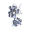
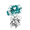
| Entry | Database: PDB / ID: 5ci6 | ||||||
|---|---|---|---|---|---|---|---|
| Title | Crystal structure of Arabidopsis thaliana MPK6 | ||||||
 Components Components | Mitogen-activated protein kinase 6 | ||||||
 Keywords Keywords |  TRANSFERASE / Kinase / apoenzyme TRANSFERASE / Kinase / apoenzyme | ||||||
| Function / homology |  Function and homology informationpreprophase band / priming of cellular response to stress / camalexin biosynthetic process / response to freezing / regulation of unidimensional cell growth / pollen tube guidance / regulation of root meristem growth / induced systemic resistance, jasmonic acid mediated signaling pathway / inflorescence development / plant ovule development ...preprophase band / priming of cellular response to stress / camalexin biosynthetic process / response to freezing / regulation of unidimensional cell growth / pollen tube guidance / regulation of root meristem growth / induced systemic resistance, jasmonic acid mediated signaling pathway / inflorescence development / plant ovule development / phragmoplast / plant-type hypersensitive response / response to ethylene / regulation of stomatal closure / leaf senescence / pollen development / root development / response to fungus / response to abscisic acid / abscisic acid-activated signaling pathway / response to UV-B / response to osmotic stress / response to L-glutamate / MAP kinase activity / mitogen-activated protein kinase / phosphatase binding / response to salt stress / response to cold / response to reactive oxygen species / trans-Golgi network / response to hydrogen peroxide / cell cortex / response to oxidative stress / protein kinase activity / intracellular signal transduction / defense response to bacterium / cell division / protein phosphorylation / protein serine kinase activity / protein serine/threonine kinase activity / ATP binding / nucleus / cytosol / cytoplasm Function and homology informationpreprophase band / priming of cellular response to stress / camalexin biosynthetic process / response to freezing / regulation of unidimensional cell growth / pollen tube guidance / regulation of root meristem growth / induced systemic resistance, jasmonic acid mediated signaling pathway / inflorescence development / plant ovule development ...preprophase band / priming of cellular response to stress / camalexin biosynthetic process / response to freezing / regulation of unidimensional cell growth / pollen tube guidance / regulation of root meristem growth / induced systemic resistance, jasmonic acid mediated signaling pathway / inflorescence development / plant ovule development / phragmoplast / plant-type hypersensitive response / response to ethylene / regulation of stomatal closure / leaf senescence / pollen development / root development / response to fungus / response to abscisic acid / abscisic acid-activated signaling pathway / response to UV-B / response to osmotic stress / response to L-glutamate / MAP kinase activity / mitogen-activated protein kinase / phosphatase binding / response to salt stress / response to cold / response to reactive oxygen species / trans-Golgi network / response to hydrogen peroxide / cell cortex / response to oxidative stress / protein kinase activity / intracellular signal transduction / defense response to bacterium / cell division / protein phosphorylation / protein serine kinase activity / protein serine/threonine kinase activity / ATP binding / nucleus / cytosol / cytoplasmSimilarity search - Function | ||||||
| Biological species |  Arabidopsis thaliana (thale cress) Arabidopsis thaliana (thale cress) | ||||||
| Method | X-RAY DIFFRACTION / SYNCHROTRON / MOLECULAR REPLACEMENT / Resolution: 3 Å | ||||||
 Authors Authors | Qin, X. / Li, P. / Chen, Z. / Ren, D. | ||||||
| Funding support |  China, 1items China, 1items
| ||||||
 Citation Citation | Journal: Sci Rep / Year: 2016 Title: Analysis of crystal structure of Arabidopsis MPK6 and generation of its mutants with higher activity Authors: Wang, B. / Qin, X. / Wu, J. / Deng, H. / Li, Y. / Yang, H. / Chen, Z. / Liu, G. / Ren, D. | ||||||
| History |
|
- Structure visualization
| Structure viewer | Molecule: MolmilJmol/JSmol |
|---|
- Downloads & links
-Download
| PDBx/mmCIF format | 5ci6.cif.gz | 148.8 KB | Display | PDBx/mmCIF format |
|---|---|---|---|---|
| PDB format | pdb5ci6.ent.gz | 116.3 KB | Display | PDB format |
| PDBx/mmJSON format | 5ci6.json.gz | Tree view | PDBx/mmJSON format | |
| Others |  Other downloads Other downloads |
-Validation report
| Arichive directory | https://data.pdbj.org/pub/pdb/validation_reports/ci/5ci6ftp://data.pdbj.org/pub/pdb/validation_reports/ci/5ci6 | HTTPS FTP |
|---|
-Related structure data
| Related structure data |  1erk S: Starting model for refinement |
|---|---|
| Similar structure data |


-Links
 PDBj
PDBj- Assembly
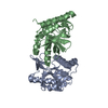
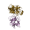
| Deposited unit | 
| ||||||||||||||||||
|---|---|---|---|---|---|---|---|---|---|---|---|---|---|---|---|---|---|---|---|
| 1 | 
| ||||||||||||||||||
| 2 | 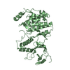
| ||||||||||||||||||
| Unit cell |
| ||||||||||||||||||
| Noncrystallographic symmetry (NCS) | NCS domain:
NCS domain segments: Component-ID: 0 / Ens-ID: 1 / Beg auth comp-ID: GLY / Beg label comp-ID: GLY / End auth comp-ID: TYR / End label comp-ID: TYR / Refine code: 0 / Auth seq-ID: 32 - 393 / Label seq-ID: 7 - 368
|
-Components
| #1: Protein | Mass: 42835.035 Da / Num. of mol.: 2 / Fragment: UNP residues 29-395 Source method: isolated from a genetically manipulated source Source: (gene. exp.) Arabidopsis thaliana (thale cress) / Gene: MPK6, At2g43790, F18O19.10 / Production host:  Escherichia coli (E. coli) Escherichia coli (E. coli)References: UniProt: Q39026, mitogen-activated protein kinase#2: Water | ChemComp-HOH / | Water Mass: 18.015 Da / Num. of mol.: 6 / Source method: isolated from a natural source / Formula: H2O Mass: 18.015 Da / Num. of mol.: 6 / Source method: isolated from a natural source / Formula: H2O |
|---|
-Experimental details
-Experiment
| Experiment | Method: X-RAY DIFFRACTION / Number of used crystals: 1 |
|---|
- Sample preparation
| Crystal | Density Matthews: 3.36 Å3/Da / Density % sol: 63.45 % |
|---|---|
| Crystal grow | Temperature: 295 K / Method: vapor diffusion, sitting drop / pH: 6.2 / Details: 0.1 M citrate, 10-12% PEG 20000 |
-Data collection
| Diffraction | Mean temperature: 100 K |
|---|---|
| Diffraction source | Source: SYNCHROTRON / Site: SSRF / Beamline: BL17U / Wavelength: 0.97923 Å |
| Detector | Type: ADSC QUANTUM 315 / Detector: CCD / Date: Jan 3, 2012 |
| Radiation | Protocol: SINGLE WAVELENGTH / Monochromatic (M) / Laue (L): M / Scattering type: x-ray |
| Radiation wavelength | Wavelength: 0.97923 Å / Relative weight: 1 |
| Reflection | Resolution: 2.9→50 Å / Num. obs: 24998 / % possible obs: 99.5 % / Redundancy: 3.6 % / Rmerge(I) obs: 0.072 / Net I/σ(I): 16.48 |
| Reflection shell | Resolution: 2.9→2.95 Å / Redundancy: 3.7 % / Rmerge(I) obs: 0.684 / Mean I/σ(I) obs: 2.15 / % possible all: 100 |
- Processing
| Software |
| ||||||||||||||||||||||||||||||||||||||||||||||||||||||||||||||||||||||||||||||||||||||||||||||||||||||||||||||||||||||||||||||||||||||||||||||||||||||||||||||||||||||||||||||||||||||
|---|---|---|---|---|---|---|---|---|---|---|---|---|---|---|---|---|---|---|---|---|---|---|---|---|---|---|---|---|---|---|---|---|---|---|---|---|---|---|---|---|---|---|---|---|---|---|---|---|---|---|---|---|---|---|---|---|---|---|---|---|---|---|---|---|---|---|---|---|---|---|---|---|---|---|---|---|---|---|---|---|---|---|---|---|---|---|---|---|---|---|---|---|---|---|---|---|---|---|---|---|---|---|---|---|---|---|---|---|---|---|---|---|---|---|---|---|---|---|---|---|---|---|---|---|---|---|---|---|---|---|---|---|---|---|---|---|---|---|---|---|---|---|---|---|---|---|---|---|---|---|---|---|---|---|---|---|---|---|---|---|---|---|---|---|---|---|---|---|---|---|---|---|---|---|---|---|---|---|---|---|---|---|---|
| Refinement | Method to determine structure: MOLECULAR REPLACEMENT Starting model: 1ERK 1erk Resolution: 3→42.7 Å / Cor.coef. Fo:Fc: 0.937 / Cor.coef. Fo:Fc free: 0.907 / SU B: 14.95 / SU ML: 0.281 / Cross valid method: THROUGHOUT / ESU R: 0.153 / ESU R Free: 0.089 / Stereochemistry target values: MAXIMUM LIKELIHOOD / Details: HYDROGENS HAVE BEEN ADDED IN THE RIDING POSITIONS
| ||||||||||||||||||||||||||||||||||||||||||||||||||||||||||||||||||||||||||||||||||||||||||||||||||||||||||||||||||||||||||||||||||||||||||||||||||||||||||||||||||||||||||||||||||||||
| Solvent computation | Ion probe radii: 0.8 Å / Shrinkage radii: 0.8 Å / VDW probe radii: 1.2 Å / Solvent model: MASK | ||||||||||||||||||||||||||||||||||||||||||||||||||||||||||||||||||||||||||||||||||||||||||||||||||||||||||||||||||||||||||||||||||||||||||||||||||||||||||||||||||||||||||||||||||||||
| Displacement parameters | Biso mean: 96.859 Å2
| ||||||||||||||||||||||||||||||||||||||||||||||||||||||||||||||||||||||||||||||||||||||||||||||||||||||||||||||||||||||||||||||||||||||||||||||||||||||||||||||||||||||||||||||||||||||
| Refinement step | Cycle: LAST / Resolution: 3→42.7 Å
| ||||||||||||||||||||||||||||||||||||||||||||||||||||||||||||||||||||||||||||||||||||||||||||||||||||||||||||||||||||||||||||||||||||||||||||||||||||||||||||||||||||||||||||||||||||||
| Refine LS restraints |
|