 Movie
Movie Controller

[English] 日本語
 Yorodumi
- PDB-5chl: Structural basis of H2A.Z recognition by YL1 histone chaperone co... -
+ Open data

- Basic information
| Entry | Database: PDB / ID: 5chl | ||||||
|---|---|---|---|---|---|---|---|
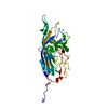
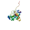
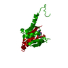
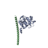
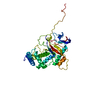
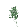
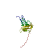
| Title | Structural basis of H2A.Z recognition by YL1 histone chaperone component of SRCAP/SWR1 chromatin remodeling complex | ||||||
 Components Components |
| ||||||
 Keywords Keywords |  CHAPERONE / remodeling complex / histone CHAPERONE / remodeling complex / histone | ||||||
| Function / homology |  Function and homology information: / : / NuA4 histone acetyltransferase complex / heterochromatin organization / RNA polymerase II core promoter sequence-specific DNA binding / nucleosomal DNA binding / heterochromatin / cellular response to estradiol stimulus / euchromatin / chromatin DNA binding ...: / : / NuA4 histone acetyltransferase complex / heterochromatin organization / RNA polymerase II core promoter sequence-specific DNA binding / nucleosomal DNA binding / heterochromatin / cellular response to estradiol stimulus / euchromatin / chromatin DNA binding / structural constituent of chromatin / nucleosome / chromatin organization / RNA polymerase II cis-regulatory region sequence-specific DNA binding / protein heterodimerization activity / Amyloid fiber formation / negative regulation of gene expression / regulation of DNA-templated transcription / positive regulation of transcription by RNA polymerase II / DNA binding / extracellular exosome / nucleus Function and homology information: / : / NuA4 histone acetyltransferase complex / heterochromatin organization / RNA polymerase II core promoter sequence-specific DNA binding / nucleosomal DNA binding / heterochromatin / cellular response to estradiol stimulus / euchromatin / chromatin DNA binding ...: / : / NuA4 histone acetyltransferase complex / heterochromatin organization / RNA polymerase II core promoter sequence-specific DNA binding / nucleosomal DNA binding / heterochromatin / cellular response to estradiol stimulus / euchromatin / chromatin DNA binding / structural constituent of chromatin / nucleosome / chromatin organization / RNA polymerase II cis-regulatory region sequence-specific DNA binding / protein heterodimerization activity / Amyloid fiber formation / negative regulation of gene expression / regulation of DNA-templated transcription / positive regulation of transcription by RNA polymerase II / DNA binding / extracellular exosome / nucleusSimilarity search - Function | ||||||
| Biological species |  Drosophila melanogaster (fruit fly) Drosophila melanogaster (fruit fly) Homo sapiens (human) Homo sapiens (human) | ||||||
| Method | X-RAY DIFFRACTION / SYNCHROTRON / Resolution: 1.892 Å | ||||||
 Authors Authors | Shan, S. / Liang, X. / Pan, L. / Wu, C. / Zhou, Z. | ||||||
 Citation Citation | Journal: Nat.Struct.Mol.Biol. / Year: 2016 Title: Structural basis of H2A.Z recognition by SRCAP chromatin-remodeling subunit YL1 Authors: Liang, X. / Shan, S. / Pan, L. / Zhao, J. / Ranjan, A. / Wang, F. / Zhang, Z. / Huang, Y. / Feng, H. / Wei, D. / Huang, L. / Liu, X. / Zhong, Q. / Lou, J. / Li, G. / Wu, C. / Zhou, Z. | ||||||
| History |
|
- Structure visualization
| Structure viewer | Molecule: MolmilJmol/JSmol |
|---|
- Downloads & links
-Download
| PDBx/mmCIF format | 5chl.cif.gz | 61.2 KB | Display | PDBx/mmCIF format |
|---|---|---|---|---|
| PDB format | pdb5chl.ent.gz | 47.3 KB | Display | PDB format |
| PDBx/mmJSON format | 5chl.json.gz | Tree view | PDBx/mmJSON format | |
| Others |  Other downloads Other downloads |
-Validation report
| Arichive directory | https://data.pdbj.org/pub/pdb/validation_reports/ch/5chlftp://data.pdbj.org/pub/pdb/validation_reports/ch/5chl | HTTPS FTP |
|---|
-Related structure data
| Similar structure data |
|---|

-Links
 PDBj
PDBj


- Assembly
| Deposited unit | 
| ||||||||
|---|---|---|---|---|---|---|---|---|---|
| 1 |
| ||||||||
| Unit cell |
| ||||||||
| Components on special symmetry positions |
|
-Components
| #1: Protein | Vacuole / Protein YL-1 Mass: 8585.671 Da / Num. of mol.: 1 Source method: isolated from a genetically manipulated source Source: (gene. exp.) Drosophila melanogaster (fruit fly) / Gene: YL-1, CG4621 / Cell line (production host): BL21(RIPL) / Production host:  Escherichia coli (E. coli) / References: UniProt: Q9VKM6 Escherichia coli (E. coli) / References: UniProt: Q9VKM6 |
|---|---|
| #2: Protein | / H2A/z Mass: 21161.436 Da / Num. of mol.: 1 Source method: isolated from a genetically manipulated source Source: (gene. exp.) Homo sapiens (human) / Gene: H2AFZ, H2AZ / Cell line (production host): BL21(RIPL) / Production host: Escherichia coli (E. coli) / References: UniProt: P0C0S5 |
| #3: Water | ChemComp-HOH / Water Mass: 18.015 Da / Num. of mol.: 147 / Source method: isolated from a natural source / Formula: H2O Mass: 18.015 Da / Num. of mol.: 147 / Source method: isolated from a natural source / Formula: H2O |
-Experimental details
-Experiment
| Experiment | Method: X-RAY DIFFRACTION |
|---|
- Sample preparation
| Crystal | Density Matthews: 2.86 Å3/Da / Density % sol: 56.95 % |
|---|---|
| Crystal grow | Temperature: 289 K / Method: vapor diffusion, hanging drop Details: 0.19 mM CYMAL-7, 0.1 M HEPES, pH 7.5, 40% Polyethylene glycol 400 |
-Data collection
| Diffraction | Mean temperature: 77 K |
|---|---|
| Diffraction source | Source: SYNCHROTRON / Site: SSRF  / Beamline: BL17U / Wavelength: 0.987 Å / Beamline: BL17U / Wavelength: 0.987 Å |
| Detector | Type: RIGAKU RAXIS IV / Detector: IMAGE PLATE / Date: Jul 7, 2013 |
| Radiation | Protocol: SINGLE WAVELENGTH / Monochromatic (M) / Laue (L): M / Scattering type: x-ray |
| Radiation wavelength | Wavelength: 0.987 Å / Relative weight: 1 |
| Reflection | Resolution: 1.892→50 Å / Num. obs: 28027 / % possible obs: 99.9 % / Redundancy: 8.8 % / Net I/σ(I): 23.3 |
- Processing
| Software |
| |||||||||||||||||||||||||||||||||||||||||||||||||||||||||||||||||||||||||||||
|---|---|---|---|---|---|---|---|---|---|---|---|---|---|---|---|---|---|---|---|---|---|---|---|---|---|---|---|---|---|---|---|---|---|---|---|---|---|---|---|---|---|---|---|---|---|---|---|---|---|---|---|---|---|---|---|---|---|---|---|---|---|---|---|---|---|---|---|---|---|---|---|---|---|---|---|---|---|---|
| Refinement | Resolution: 1.892→29.967 Å / SU ML: 0.14 / Cross valid method: FREE R-VALUE / σ(F): 1.36 / Phase error: 22.8 / Stereochemistry target values: ML
| |||||||||||||||||||||||||||||||||||||||||||||||||||||||||||||||||||||||||||||
| Solvent computation | Shrinkage radii: 0.9 Å / VDW probe radii: 1.11 Å / Solvent model: FLAT BULK SOLVENT MODEL | |||||||||||||||||||||||||||||||||||||||||||||||||||||||||||||||||||||||||||||
| Refinement step | Cycle: LAST / Resolution: 1.892→29.967 Å
| |||||||||||||||||||||||||||||||||||||||||||||||||||||||||||||||||||||||||||||
| Refine LS restraints |
| |||||||||||||||||||||||||||||||||||||||||||||||||||||||||||||||||||||||||||||
| LS refinement shell |
|
