 Movie
Movie Controller

[English] 日本語
 Yorodumi
- PDB-5cey: Crystal Structure of Fab 9H+3L, a putative precursor of the PGT12... -
+ Open data

- Basic information
| Entry | Database: PDB / ID: 5cey | ||||||
|---|---|---|---|---|---|---|---|
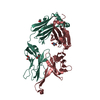
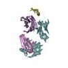
| Title | Crystal Structure of Fab 9H+3L, a putative precursor of the PGT121 family of potent HIV-1 antibodies | ||||||
 Components Components | (Antibody 9H+3L Fab ...) x 2 | ||||||
 Keywords Keywords |  IMMUNE SYSTEM / HIV-1 / antibody IMMUNE SYSTEM / HIV-1 / antibody | ||||||
| Function / homology | Immunoglobulins / Immunoglobulin-like / Sandwich / Mainly Beta / 1-DEOXY-1-THIO-HEPTAETHYLENE GLYCOL Function and homology information Function and homology information | ||||||
| Biological species |  Homo sapiens (human) Homo sapiens (human) | ||||||
| Method | X-RAY DIFFRACTION / SYNCHROTRON / MOLECULAR REPLACEMENT / Resolution: 2.387 Å | ||||||
 Authors Authors | Wilson, I.A. / Garces, F. | ||||||
 Citation Citation | Journal: Immunity / Year: 2015 Title: Affinity Maturation of a Potent Family of HIV Antibodies Is Primarily Focused on Accommodating or Avoiding Glycans. Authors: Fernando Garces / Jeong Hyun Lee / Natalia de Val / Alba Torrents de la Pena / Leopold Kong / Cristina Puchades / Yuanzi Hua / Robyn L Stanfield / Dennis R Burton / John P Moore / Rogier W ...Authors: Fernando Garces / Jeong Hyun Lee / Natalia de Val / Alba Torrents de la Pena / Leopold Kong / Cristina Puchades / Yuanzi Hua / Robyn L Stanfield / Dennis R Burton / John P Moore / Rogier W Sanders / Andrew B Ward / Ian A Wilson /   Abstract: The high-mannose patch on the HIV-1 envelope (Env) glycoprotein is the epicenter for binding of the potent broadly neutralizing PGT121 family of antibodies, but strategies for generating such ...The high-mannose patch on the HIV-1 envelope (Env) glycoprotein is the epicenter for binding of the potent broadly neutralizing PGT121 family of antibodies, but strategies for generating such antibodies by vaccination have not been defined. We generated structures of inferred antibody intermediates by X-ray crystallography and electron microscopy to elucidate the molecular events that occurred during evolution of this family. Binding analyses revealed that affinity maturation was primarily focused on avoiding, accommodating, or binding the N137 glycan. The overall antibody approach angle to Env was defined very early in the maturation process, yet some variation evolved in the PGT121 family branches that led to differences in glycan specificities in their respective epitopes. Furthermore, we determined a crystal structure of the recombinant BG505 SOSIP.664 HIV-1 trimer with a PGT121 family member at 3.0 Å that, in concert with these antibody intermediate structures, provides insights to advance design of HIV vaccine candidates. | ||||||
| History |
|
- Structure visualization
| Structure viewer | Molecule: MolmilJmol/JSmol |
|---|
- Downloads & links
-Download
| PDBx/mmCIF format | 5cey.cif.gz | 181.8 KB | Display | PDBx/mmCIF format |
|---|---|---|---|---|
| PDB format | pdb5cey.ent.gz | 142.3 KB | Display | PDB format |
| PDBx/mmJSON format | 5cey.json.gz | Tree view | PDBx/mmJSON format | |
| Others |  Other downloads Other downloads |
-Validation report
| Arichive directory | https://data.pdbj.org/pub/pdb/validation_reports/ce/5ceyftp://data.pdbj.org/pub/pdb/validation_reports/ce/5cey | HTTPS FTP |
|---|
-Related structure data
| Related structure data |  3092C  3093C  6379C  6380C  5cexC  5cezC  4r26S C: citing same article ( S: Starting model for refinement |
|---|---|
| Similar structure data |


-Links
 PDBj
PDBj


- Assembly
| Deposited unit | 
| ||||||||
|---|---|---|---|---|---|---|---|---|---|
| 1 | 
| ||||||||
| 2 | 
| ||||||||
| Unit cell |
|
-Components
-Antibody , 2 types, 4 molecules ACBD
| #1: Antibody | Mass: 23426.996 Da / Num. of mol.: 2 Source method: isolated from a genetically manipulated source Source: (gene. exp.) Homo sapiens (human) / Production host: Homo sapiens (human)#2: Antibody | Mass: 25419.559 Da / Num. of mol.: 2 Source method: isolated from a genetically manipulated source Source: (gene. exp.) Homo sapiens (human) / Production host: Homo sapiens (human) |
|---|
-Non-polymers , 4 types, 106 molecules 


| #3: Chemical | Glycerol Mass: 92.094 Da / Num. of mol.: 2 / Source method: obtained synthetically / Formula: C3H8O3 Mass: 92.094 Da / Num. of mol.: 2 / Source method: obtained synthetically / Formula: C3H8O3#4: Chemical | ChemComp-P6G / | Polyethylene glycol Mass: 282.331 Da / Num. of mol.: 1 / Source method: obtained synthetically / Formula: C12H26O7 / Comment: precipitant*YM Mass: 282.331 Da / Num. of mol.: 1 / Source method: obtained synthetically / Formula: C12H26O7 / Comment: precipitant*YM#5: Chemical | ChemComp-PE7 / |  Mass: 342.449 Da / Num. of mol.: 1 / Source method: obtained synthetically / Formula: C14H30O7S Mass: 342.449 Da / Num. of mol.: 1 / Source method: obtained synthetically / Formula: C14H30O7S#6: Water | ChemComp-HOH / | WaterMass: 18.015 Da / Num. of mol.: 102 / Source method: isolated from a natural source / Formula: H2O |
|---|
-Experimental details
-Experiment
| Experiment | Method: X-RAY DIFFRACTION / Number of used crystals: 1 |
|---|
- Sample preparation
| Crystal | Density Matthews: 2.23 Å3/Da / Density % sol: 44.89 % |
|---|---|
| Crystal grow | Temperature: 293.14 K / Method: vapor diffusion, sitting drop Details: 40% (v/v) PEG 600, 0.1M NaCl and 0.1 M Na citrate pH 5.5 |
-Data collection
| Diffraction | Mean temperature: 100 K |
|---|---|
| Diffraction source | Source: SYNCHROTRON / Site: APS / Beamline: 21-ID-D / Wavelength: 1.033 Å |
| Detector | Type: PSI PILATUS 6M / Detector: PIXEL / Date: Oct 25, 2014 |
| Radiation | Protocol: SINGLE WAVELENGTH / Monochromatic (M) / Laue (L): M / Scattering type: x-ray |
| Radiation wavelength | Wavelength: 1.033 Å / Relative weight: 1 |
| Reflection | Resolution: 2.387→45.747 Å / Num. obs: 33105 / % possible obs: 94.2 % / Redundancy: 4.6 % / Rsym value: 0.13 / Net I/σ(I): 7.87 |
| Reflection shell | Highest resolution: 2.4 Å / Redundancy: 3.9 % / Mean I/σ(I) obs: 4.7 / Rsym value: 0.26 / % possible all: 72.2 |
- Processing
| Software |
| |||||||||||||||||||||||||||||||||||||||||||||||||||||||||||||||||||||||||||||||||||||||||||
|---|---|---|---|---|---|---|---|---|---|---|---|---|---|---|---|---|---|---|---|---|---|---|---|---|---|---|---|---|---|---|---|---|---|---|---|---|---|---|---|---|---|---|---|---|---|---|---|---|---|---|---|---|---|---|---|---|---|---|---|---|---|---|---|---|---|---|---|---|---|---|---|---|---|---|---|---|---|---|---|---|---|---|---|---|---|---|---|---|---|---|---|---|
| Refinement | Method to determine structure: MOLECULAR REPLACEMENT Starting model: 4R26 Resolution: 2.387→45.747 Å / SU ML: 0.33 / Cross valid method: NONE / σ(F): 1.34 / Phase error: 30.11 / Stereochemistry target values: ML
| |||||||||||||||||||||||||||||||||||||||||||||||||||||||||||||||||||||||||||||||||||||||||||
| Solvent computation | Shrinkage radii: 0.9 Å / VDW probe radii: 1.11 Å / Solvent model: FLAT BULK SOLVENT MODEL | |||||||||||||||||||||||||||||||||||||||||||||||||||||||||||||||||||||||||||||||||||||||||||
| Refinement step | Cycle: LAST / Resolution: 2.387→45.747 Å
| |||||||||||||||||||||||||||||||||||||||||||||||||||||||||||||||||||||||||||||||||||||||||||
| Refine LS restraints |
| |||||||||||||||||||||||||||||||||||||||||||||||||||||||||||||||||||||||||||||||||||||||||||
| LS refinement shell |
|
