 Movie
Movie Controller


+ Open data

- Basic information
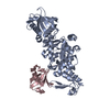
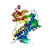
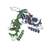
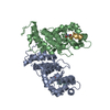
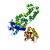
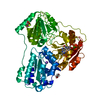
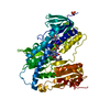
| Entry | Database: PDB / ID: 5c91 | ||||||
|---|---|---|---|---|---|---|---|
| Title | NEDD4 HECT with covalently bound indole-based inhibitor | ||||||
 Components Components | E3 ubiquitin-protein ligase NEDD4 | ||||||
 Keywords Keywords | ligase/ligase inhibitor /  NEDD4 / HECT / LIGASE / INHIBITOR / ligase-ligase inhibitor complex NEDD4 / HECT / LIGASE / INHIBITOR / ligase-ligase inhibitor complex | ||||||
| Function / homology |  Function and homology information Function and homology informationformation of structure involved in a symbiotic process / positive regulation of nucleocytoplasmic transport / negative regulation of sodium ion transport / regulation of potassium ion transmembrane transporter activity / viral budding / intracellular glucocorticoid receptor signaling pathway / negative regulation of sodium ion transmembrane transporter activity / phosphothreonine residue binding / receptor catabolic process / protein targeting to lysosome ...formation of structure involved in a symbiotic process / positive regulation of nucleocytoplasmic transport / negative regulation of sodium ion transport / regulation of potassium ion transmembrane transporter activity / viral budding / intracellular glucocorticoid receptor signaling pathway / negative regulation of sodium ion transmembrane transporter activity / phosphothreonine residue binding / receptor catabolic process / protein targeting to lysosome / apicolateral plasma membrane / ubiquitin-dependent protein catabolic process via the multivesicular body sorting pathway / HECT-type E3 ubiquitin transferase / sodium channel inhibitor activity / proline-rich region binding / regulation of monoatomic ion transmembrane transport / RNA polymerase binding / lysosomal transport / beta-2 adrenergic receptor binding / neuromuscular junction development / regulation of dendrite morphogenesis / regulation of synapse organization / negative regulation of vascular endothelial growth factor receptor signaling pathway / progesterone receptor signaling pathway / protein K63-linked ubiquitination / phosphoserine residue binding / regulation of macroautophagy / ubiquitin ligase complex / Downregulation of ERBB4 signaling / Regulation of PTEN localization / ubiquitin binding / regulation of membrane potential / receptor internalization / ISG15 antiviral mechanism / Regulation of PTEN stability and activity / response to calcium ion / positive regulation of protein catabolic process / cellular response to UV / neuron projection development / ubiquitin protein ligase activity / Antigen processing: Ubiquitination & Proteasome degradation / cell cortex / ubiquitin-dependent protein catabolic process / dendritic spine / positive regulation of phosphatidylinositol 3-kinase/protein kinase B signal transduction / protein ubiquitination / protein domain specific binding / innate immune response / DNA damage response / chromatin / perinuclear region of cytoplasm / Golgi apparatus / enzyme binding / negative regulation of transcription by RNA polymerase II / protein-containing complex / extracellular exosome / nucleus / plasma membrane / cytosol / cytoplasmSimilarity search - Function | ||||||
| Biological species |  Homo sapiens (human) Homo sapiens (human) | ||||||
| Method | X-RAY DIFFRACTION / SYNCHROTRON / MOLECULAR REPLACEMENT / Resolution: 2.44 Å | ||||||
 Authors Authors | Span, I. / Smith, A.T. / Kathman, S. / Statsyuk, A.V. / Rosenzweig, A.C. | ||||||
 Citation Citation | Journal: J. Am. Chem. Soc. / Year: 2015 Title: A Small Molecule That Switches a Ubiquitin Ligase From a Processive to a Distributive Enzymatic Mechanism. Authors: Kathman, S.G. / Span, I. / Smith, A.T. / Xu, Z. / Zhan, J. / Rosenzweig, A.C. / Statsyuk, A.V. | ||||||
| History |
|
- Structure visualization
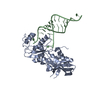
| Structure viewer | Molecule: MolmilJmol/JSmol |
|---|
- Downloads & links
-Download
| PDBx/mmCIF format | 5c91.cif.gz | 166.7 KB | Display | PDBx/mmCIF format |
|---|---|---|---|---|
| PDB format | pdb5c91.ent.gz | 130.4 KB | Display | PDB format |
| PDBx/mmJSON format | 5c91.json.gz | Tree view | PDBx/mmJSON format | |
| Others |  Other downloads Other downloads |
-Validation report
| Arichive directory | https://data.pdbj.org/pub/pdb/validation_reports/c9/5c91ftp://data.pdbj.org/pub/pdb/validation_reports/c9/5c91 | HTTPS FTP |
|---|
-Related structure data
| Related structure data |  2xbfS S: Starting model for refinement |
|---|---|
| Similar structure data |


-Links
 PDBj
PDBj


- Assembly
| Deposited unit | 
| ||||||||
|---|---|---|---|---|---|---|---|---|---|
| 1 |
| ||||||||
| Unit cell |
|
-Components
| #1: Protein | Mass: 44739.031 Da / Num. of mol.: 1 / Fragment: unp residues 938-1312 Source method: isolated from a genetically manipulated source Source: (gene. exp.) Homo sapiens (human) / Gene: NEDD4, KIAA0093, NEDD4-1, PIG53 / Production host:  Escherichia coli (E. coli) Escherichia coli (E. coli)References: UniProt: P46934, Ligases; Forming carbon-nitrogen bonds; Acid-amino-acid ligases (peptide synthases) |
|---|---|
| #2: Chemical | ChemComp-4YU /   Mass: 316.352 Da / Num. of mol.: 1 / Source method: obtained synthetically / Formula: C17H20N2O4 Mass: 316.352 Da / Num. of mol.: 1 / Source method: obtained synthetically / Formula: C17H20N2O4 |
| #3: Water | ChemComp-HOH / Water Mass: 18.015 Da / Num. of mol.: 7 / Source method: isolated from a natural source / Formula: H2O Mass: 18.015 Da / Num. of mol.: 7 / Source method: isolated from a natural source / Formula: H2O |
-Experimental details
-Experiment
| Experiment | Method: X-RAY DIFFRACTION / Number of used crystals: 1 |
|---|
- Sample preparation
| Crystal | Density Matthews: 2.29 Å3/Da / Density % sol: 46.18 % |
|---|---|
| Crystal grow | Temperature: 293 K / Method: vapor diffusion, hanging drop Details: 100 mM MES, 35 mM calcium chloride, 5 mM TCEP, 6% PEG400 PH range: 6.5 |
-Data collection
| Diffraction | Mean temperature: 100 K | |||||||||||||||||||||
|---|---|---|---|---|---|---|---|---|---|---|---|---|---|---|---|---|---|---|---|---|---|---|
| Diffraction source | Source: SYNCHROTRON / Site: APS  / Beamline: 23-ID-D / Wavelength: 1.033 Å / Beamline: 23-ID-D / Wavelength: 1.033 Å | |||||||||||||||||||||
| Detector | Type: PSI PILATUS 6M / Detector: PIXEL / Date: Mar 10, 2014 | |||||||||||||||||||||
| Radiation | Monochromator: Double crystal cryo-cooled / Protocol: SINGLE WAVELENGTH / Monochromatic (M) / Laue (L): M / Scattering type: x-ray | |||||||||||||||||||||
| Radiation wavelength | Wavelength: 1.033 Å / Relative weight: 1 | |||||||||||||||||||||
| Reflection | Resolution: 2.44→32.2 Å / Num. obs: 15105 / % possible obs: 98.2 % / Redundancy: 3.2 % / Biso Wilson estimate: 23.03 Å2 / Rmerge(I) obs: 0.091 / Rpim(I) all: 0.071 / Net I/σ(I): 11.2 / Num. measured all: 47656 | |||||||||||||||||||||
| Reflection shell | Diffraction-ID: 1 / Redundancy: 3.1 % / Rejects: 0
|
- Processing
| Software |
| |||||||||||||||||||||||||||||||||||||||||||||||||||||||||||||||||||||||||||
|---|---|---|---|---|---|---|---|---|---|---|---|---|---|---|---|---|---|---|---|---|---|---|---|---|---|---|---|---|---|---|---|---|---|---|---|---|---|---|---|---|---|---|---|---|---|---|---|---|---|---|---|---|---|---|---|---|---|---|---|---|---|---|---|---|---|---|---|---|---|---|---|---|---|---|---|---|
| Refinement | Method to determine structure: MOLECULAR REPLACEMENT Starting model: 2XBF Resolution: 2.44→31.86 Å / Cor.coef. Fo:Fc: 0.918 / Cor.coef. Fo:Fc free: 0.877 / SU B: 36.163 / SU ML: 0.359 / Cross valid method: THROUGHOUT / σ(F): 0 / ESU R: 0.89 / ESU R Free: 0.351 / Stereochemistry target values: MAXIMUM LIKELIHOOD Details: HYDROGENS HAVE BEEN ADDED IN THE RIDING POSITIONS U VALUES : WITH TLS ADDED
| |||||||||||||||||||||||||||||||||||||||||||||||||||||||||||||||||||||||||||
| Solvent computation | Ion probe radii: 0.8 Å / Shrinkage radii: 0.8 Å / VDW probe radii: 1.2 Å / Solvent model: MASK | |||||||||||||||||||||||||||||||||||||||||||||||||||||||||||||||||||||||||||
| Displacement parameters | Biso max: 111.45 Å2 / Biso mean: 50.376 Å2 / Biso min: 24.6 Å2
| |||||||||||||||||||||||||||||||||||||||||||||||||||||||||||||||||||||||||||
| Refinement step | Cycle: final / Resolution: 2.44→31.86 Å
| |||||||||||||||||||||||||||||||||||||||||||||||||||||||||||||||||||||||||||
| Refine LS restraints |
| |||||||||||||||||||||||||||||||||||||||||||||||||||||||||||||||||||||||||||
| LS refinement shell | Resolution: 2.44→2.503 Å / Total num. of bins used: 20
| |||||||||||||||||||||||||||||||||||||||||||||||||||||||||||||||||||||||||||
| Refinement TLS params. | Method: refined / Details: Chain A / Origin x: -22.8336 Å / Origin y: -2.3324 Å / Origin z: 8.9346 Å
|
