 Movie
Movie Controller

[English] 日本語
 Yorodumi
- PDB-5c8f: Crystal structure of light-exposed full-length Thermus thermophil... -
+ Open data

- Basic information
| Entry | Database: PDB / ID: 5c8f | |||||||||
|---|---|---|---|---|---|---|---|---|---|---|
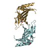
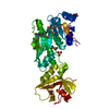
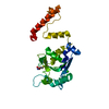
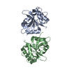
| Title | Crystal structure of light-exposed full-length Thermus thermophilus CarH bound to cobalamin | |||||||||
 Components Components | Light-dependent transcriptional regulator CarH | |||||||||
 Keywords Keywords |  TRANSCRIPTIONAL REGULATOR / Transcription factor / light sensor / adenosylcobalamin-binding / DNA-binding / TRANSCRIPTION TRANSCRIPTIONAL REGULATOR / Transcription factor / light sensor / adenosylcobalamin-binding / DNA-binding / TRANSCRIPTION | |||||||||
| Function / homology |  Function and homology informationcobalamin binding / DNA-binding transcription factor activity / DNA binding / identical protein binding / metal ion binding Function and homology informationcobalamin binding / DNA-binding transcription factor activity / DNA binding / identical protein binding / metal ion bindingSimilarity search - Function | |||||||||
| Biological species |   Thermus thermophilus (bacteria) Thermus thermophilus (bacteria) | |||||||||
| Method | X-RAY DIFFRACTION / SYNCHROTRON / MOLECULAR REPLACEMENT / Resolution: 2.65 Å | |||||||||
 Authors Authors | Jost, M. / Drennan, C.L. | |||||||||
| Funding support |  United States, 2items United States, 2items
| |||||||||
 Citation Citation | Journal: Nature / Year: 2015 Title: Structural basis for gene regulation by a B12-dependent photoreceptor. Authors: Jost, M. / Fernandez-Zapata, J. / Polanco, M.C. / Ortiz-Guerrero, J.M. / Chen, P.Y. / Kang, G. / Padmanabhan, S. / Elias-Arnanz, M. / Drennan, C.L. | |||||||||
| History |
|
- Structure visualization
| Structure viewer | Molecule: MolmilJmol/JSmol |
|---|
- Downloads & links
-Download
| PDBx/mmCIF format | 5c8f.cif.gz | 129.4 KB | Display | PDBx/mmCIF format |
|---|---|---|---|---|
| PDB format | pdb5c8f.ent.gz | 99.5 KB | Display | PDB format |
| PDBx/mmJSON format | 5c8f.json.gz | Tree view | PDBx/mmJSON format | |
| Others |  Other downloads Other downloads |
-Validation report
| Arichive directory | https://data.pdbj.org/pub/pdb/validation_reports/c8/5c8fftp://data.pdbj.org/pub/pdb/validation_reports/c8/5c8f | HTTPS FTP |
|---|
-Related structure data
| Related structure data |  5c8aC 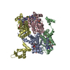 5c8dC 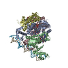 5c8eC 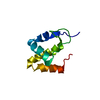 2jmlS C: citing same article ( S: Starting model for refinement |
|---|---|
| Similar structure data |


-Links
 PDBj
PDBj

- Assembly
| Deposited unit | 
| ||||||||
|---|---|---|---|---|---|---|---|---|---|
| 1 |
| ||||||||
| 2 | 
| ||||||||
| Unit cell |
| ||||||||
| Details | Monomer assembly has been verified by size exclusion chromatography and analytical ultracentrifugation. Importantly, only this particular form of the CarH protein (light-exposed CarH) is monomeric. |
-Components
| #1: Protein | Mass: 33323.250 Da / Num. of mol.: 1 Source method: isolated from a genetically manipulated source Source: (gene. exp.) Thermus thermophilus (strain HB27 / ATCC BAA-163 / DSM 7039) (bacteria)Strain: HB27 / ATCC BAA-163 / DSM 7039 / Gene: TT_P0056 / Plasmid: pET15b / Production host: Escherichia coli (E. coli) / Strain (production host): BL21 Star pLysS / References: UniProt: Q746J7 | ||||
|---|---|---|---|---|---|
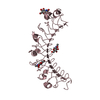
| #2: Chemical | ChemComp-B12 / Vitamin B12  Mass: 1330.356 Da / Num. of mol.: 1 / Source method: obtained synthetically / Formula: C62H89CoN13O14P Mass: 1330.356 Da / Num. of mol.: 1 / Source method: obtained synthetically / Formula: C62H89CoN13O14P | ||||
| #3: Chemical | Chloride  Mass: 35.453 Da / Num. of mol.: 2 / Source method: obtained synthetically / Formula: Cl Mass: 35.453 Da / Num. of mol.: 2 / Source method: obtained synthetically / Formula: Cl#4: Chemical | ChemComp-GOL / | Glycerol  Mass: 92.094 Da / Num. of mol.: 1 / Source method: obtained synthetically / Formula: C3H8O3 Mass: 92.094 Da / Num. of mol.: 1 / Source method: obtained synthetically / Formula: C3H8O3#5: Water | ChemComp-HOH / | Water Mass: 18.015 Da / Num. of mol.: 19 / Source method: isolated from a natural source / Formula: H2O Mass: 18.015 Da / Num. of mol.: 19 / Source method: isolated from a natural source / Formula: H2O |
-Experimental details
-Experiment
| Experiment | Method: X-RAY DIFFRACTION |
|---|
- Sample preparation
| Crystal | Density Matthews: 4.52 Å3/Da / Density % sol: 72.77 % |
|---|---|
| Crystal grow | Temperature: 298 K / Method: vapor diffusion, hanging drop / pH: 6 / Details: 3.4 M NaCl, 0.1 M Bis-Tris pH 6 |
-Data collection
| Diffraction | Mean temperature: 100 K |
|---|---|
| Diffraction source | Source: SYNCHROTRON / Site: APS / Beamline: 24-ID-C / Wavelength: 0.9791 Å |
| Detector | Type: DECTRIS PILATUS 6M-F / Detector: PIXEL / Date: Nov 23, 2014 |
| Radiation | Monochromator: Si(111) / Protocol: SINGLE WAVELENGTH / Monochromatic (M) / Laue (L): M / Scattering type: x-ray |
| Radiation wavelength | Wavelength: 0.9791 Å / Relative weight: 1 |
| Reflection | Resolution: 2.65→100 Å / Num. obs: 18067 / % possible obs: 100 % / Redundancy: 12.8 % / Rsym value: 0.069 / Net I/σ(I): 26.8 |
| Reflection shell | Resolution: 2.65→2.72 Å / Redundancy: 13.4 % / Mean I/σ(I) obs: 1.8 / % possible all: 99.9 |
- Processing
| Software |
| |||||||||||||||||||||||||||||||||||||||||||||||||
|---|---|---|---|---|---|---|---|---|---|---|---|---|---|---|---|---|---|---|---|---|---|---|---|---|---|---|---|---|---|---|---|---|---|---|---|---|---|---|---|---|---|---|---|---|---|---|---|---|---|---|
| Refinement | Method to determine structure: MOLECULAR REPLACEMENT Starting model: 2JML Resolution: 2.65→89.74 Å / SU ML: 0.21 / Cross valid method: FREE R-VALUE / Phase error: 23.16 / Stereochemistry target values: ML
| |||||||||||||||||||||||||||||||||||||||||||||||||
| Solvent computation | Shrinkage radii: 0.9 Å / VDW probe radii: 1.11 Å / Solvent model: FLAT BULK SOLVENT MODEL | |||||||||||||||||||||||||||||||||||||||||||||||||
| Refinement step | Cycle: LAST / Resolution: 2.65→89.74 Å
| |||||||||||||||||||||||||||||||||||||||||||||||||
| Refine LS restraints |
| |||||||||||||||||||||||||||||||||||||||||||||||||
| LS refinement shell |
| |||||||||||||||||||||||||||||||||||||||||||||||||
| Refinement TLS params. | Method: refined / Origin x: 6.786 Å / Origin y: 4.4298 Å / Origin z: -52.9519 Å
| |||||||||||||||||||||||||||||||||||||||||||||||||
| Refinement TLS group | Selection details: (chain 'A' ) |
