 Movie
Movie Controller


+ Open data

- Basic information
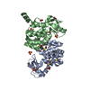
| Entry | Database: PDB / ID: 5an3 | ||||||
|---|---|---|---|---|---|---|---|
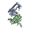
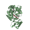
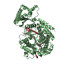
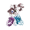
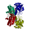
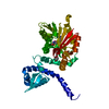
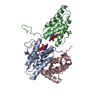
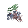
| Title | Structure of an Sgt1-Skp1 Complex | ||||||
 Components Components |
| ||||||
 Keywords Keywords |  TRANSCRIPTION TRANSCRIPTION | ||||||
| Function / homology |  Function and homology information Function and homology informationprotein-containing complex assembly => GO:0065003 / RAVE complex / Iron uptake and transport / CBF3 complex / regulation of transcription by galactose / regulation of sulfur amino acid metabolic process / cellular response to methylmercury / vacuolar proton-transporting V-type ATPase complex assembly / FBXL7 down-regulates AURKA during mitotic entry and in early mitosis / septin ring assembly ...protein-containing complex assembly => GO:0065003 / RAVE complex / Iron uptake and transport / CBF3 complex / regulation of transcription by galactose / regulation of sulfur amino acid metabolic process / cellular response to methylmercury / vacuolar proton-transporting V-type ATPase complex assembly / FBXL7 down-regulates AURKA during mitotic entry and in early mitosis / septin ring assembly / regulation of exit from mitosis / Antigen processing: Ubiquitination & Proteasome degradation / vacuolar acidification / kinetochore assembly / regulation of metabolic process / exit from mitosis / positive regulation of glucose transmembrane transport / protein neddylation / mitotic intra-S DNA damage checkpoint signaling / mitochondrial fusion / silent mating-type cassette heterochromatin formation / SCF-dependent proteasomal ubiquitin-dependent protein catabolic process / SCF ubiquitin ligase complex / DNA replication origin binding / cullin family protein binding / subtelomeric heterochromatin formation / cAMP-mediated signaling / regulation of protein-containing complex assembly / endomembrane system / negative regulation of cytoplasmic translation / ubiquitin ligase complex / regulation of mitotic cell cycle / G1/S transition of mitotic cell cycle / regulation of protein stability / kinetochore / G2/M transition of mitotic cell cycle / protein-macromolecule adaptor activity / mitotic cell cycle / ubiquitin-dependent protein catabolic process / protein-folding chaperone binding / protein-containing complex assembly / chromosome, telomeric region / protein stabilization / protein ubiquitination / cell cycle / nucleus / cytoplasmSimilarity search - Function | ||||||
| Biological species |  SACCHAROMYCES CEREVISIAE (brewer's yeast) SACCHAROMYCES CEREVISIAE (brewer's yeast) | ||||||
| Method | X-RAY DIFFRACTION / SYNCHROTRON / SAD / Resolution: 2.82 Å | ||||||
 Authors Authors | Willhoft, O. / Vaughan, C.K. | ||||||
 Citation Citation | Journal: Sci Rep / Year: 2017 Title: The crystal structure of the Sgt1-Skp1 complex: the link between Hsp90 and both SCF E3 ubiquitin ligases and kinetochores. Authors: Willhoft, O. / Kerr, R. / Patel, D. / Zhang, W. / Al-Jassar, C. / Daviter, T. / Millson, S.H. / Thalassinos, K. / Vaughan, C.K. | ||||||
| History |
|
- Structure visualization
| Structure viewer | Molecule: MolmilJmol/JSmol |
|---|
- Downloads & links
-Download
| PDBx/mmCIF format | 5an3.cif.gz | 200.3 KB | Display | PDBx/mmCIF format |
|---|---|---|---|---|
| PDB format | pdb5an3.ent.gz | 161.5 KB | Display | PDB format |
| PDBx/mmJSON format | 5an3.json.gz | Tree view | PDBx/mmJSON format | |
| Others |  Other downloads Other downloads |
-Validation report
| Arichive directory | https://data.pdbj.org/pub/pdb/validation_reports/an/5an3ftp://data.pdbj.org/pub/pdb/validation_reports/an/5an3 | HTTPS FTP |
|---|
-Related structure data
| Similar structure data |
|---|

-Links
 PDBj
PDBj


- Assembly
| Deposited unit | 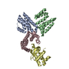
| ||||||||
|---|---|---|---|---|---|---|---|---|---|
| 1 | 
| ||||||||
| 2 | 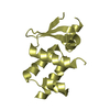
| ||||||||
| Unit cell |
|
-Components
| #1: Protein | Mass: 17492.070 Da / Num. of mol.: 3 / Fragment: TPR DOMAIN, UNP RESIDUES 1-150 Source method: isolated from a genetically manipulated source Source: (gene. exp.) SACCHAROMYCES CEREVISIAE (brewer's yeast)Production host:  ESCHERICHIA COLI (E. coli) / Strain (production host): BL21 / Variant (production host): T7 EXPRESS LYSY/IQ / References: UniProt: Q08446 ESCHERICHIA COLI (E. coli) / Strain (production host): BL21 / Variant (production host): T7 EXPRESS LYSY/IQ / References: UniProt: Q08446#2: Protein | | Mass: 14886.524 Da / Num. of mol.: 1 / Fragment: BTBPOZ DOMAIN, UNP RESIDUES 1-35,65-158 Source method: isolated from a genetically manipulated source Source: (gene. exp.) SACCHAROMYCES CEREVISIAE (brewer's yeast)Production host: ESCHERICHIA COLI (E. coli) / Strain (production host): BL21 / Variant (production host): T7 EXPRESS LYSY/IQ / References: UniProt: P52286#3: Water | ChemComp-HOH / | Water Mass: 18.015 Da / Num. of mol.: 22 / Source method: isolated from a natural source / Formula: H2O Mass: 18.015 Da / Num. of mol.: 22 / Source method: isolated from a natural source / Formula: H2O |
|---|
-Experimental details
-Experiment
| Experiment | Method: X-RAY DIFFRACTION |
|---|
- Sample preparation
| Crystal | Density Matthews: 2.72 Å3/Da / Density % sol: 54.84 % / Description: NONE |
|---|---|
| Crystal grow | Details: 0.325 M MGCL2, 22.5% PEG-6000, 0.1 M TRIS-HCL PH 8 |
-Data collection
| Diffraction | Mean temperature: 100 K |
|---|---|
| Diffraction source | Source: SYNCHROTRON / Site: Diamond  / Beamline: I04 / Wavelength: 0.979494 / Beamline: I04 / Wavelength: 0.979494 |
| Detector | Type: ADSC ADSC Q105 / Detector: CCD |
| Radiation | Protocol: SINGLE WAVELENGTH / Monochromatic (M) / Laue (L): M / Scattering type: x-ray |
| Radiation wavelength | Wavelength: 0.979494 Å / Relative weight: 1 |
| Reflection | Resolution: 2.82→81.89 Å / Num. obs: 15826 / % possible obs: 100 % / Observed criterion σ(I): -3 / Redundancy: 21.5 % / Biso Wilson estimate: 86.92 Å2 / Rmerge(I) obs: 0.09 / Net I/σ(I): 29.2 |
| Reflection shell | Resolution: 2.82→2.89 Å / Redundancy: 21.9 % / Rmerge(I) obs: 0.83 / Mean I/σ(I) obs: 4.9 / % possible all: 100 |
- Processing
| Software |
| |||||||||||||||||||||||||||||||||||||||||||||||||||||||||||||||||||||||||||||||||||||||||||||||||||||||||||||||||||||||||||||
|---|---|---|---|---|---|---|---|---|---|---|---|---|---|---|---|---|---|---|---|---|---|---|---|---|---|---|---|---|---|---|---|---|---|---|---|---|---|---|---|---|---|---|---|---|---|---|---|---|---|---|---|---|---|---|---|---|---|---|---|---|---|---|---|---|---|---|---|---|---|---|---|---|---|---|---|---|---|---|---|---|---|---|---|---|---|---|---|---|---|---|---|---|---|---|---|---|---|---|---|---|---|---|---|---|---|---|---|---|---|---|---|---|---|---|---|---|---|---|---|---|---|---|---|---|---|---|
| Refinement | Method to determine structure: SAD Starting model: NONE Resolution: 2.82→19.88 Å / Cor.coef. Fo:Fc: 0.9337 / Cor.coef. Fo:Fc free: 0.8998 / SU R Cruickshank DPI: 1.211 / Cross valid method: THROUGHOUT / σ(F): 0 / SU R Blow DPI: 1.105 / SU Rfree Blow DPI: 0.322 / SU Rfree Cruickshank DPI: 0.33 Details: IDEAL-DIST CONTACT TERM CONTACT SETUP. ALL ATOMS HAVE CCP4 ATOM TYPE FROM LIBRARY
| |||||||||||||||||||||||||||||||||||||||||||||||||||||||||||||||||||||||||||||||||||||||||||||||||||||||||||||||||||||||||||||
| Displacement parameters | Biso mean: 70.85 Å2
| |||||||||||||||||||||||||||||||||||||||||||||||||||||||||||||||||||||||||||||||||||||||||||||||||||||||||||||||||||||||||||||
| Refine analyze | Luzzati coordinate error obs: 0.441 Å | |||||||||||||||||||||||||||||||||||||||||||||||||||||||||||||||||||||||||||||||||||||||||||||||||||||||||||||||||||||||||||||
| Refinement step | Cycle: LAST / Resolution: 2.82→19.88 Å
| |||||||||||||||||||||||||||||||||||||||||||||||||||||||||||||||||||||||||||||||||||||||||||||||||||||||||||||||||||||||||||||
| Refine LS restraints |
| |||||||||||||||||||||||||||||||||||||||||||||||||||||||||||||||||||||||||||||||||||||||||||||||||||||||||||||||||||||||||||||
| LS refinement shell | Resolution: 2.82→3.02 Å / Total num. of bins used: 8
| |||||||||||||||||||||||||||||||||||||||||||||||||||||||||||||||||||||||||||||||||||||||||||||||||||||||||||||||||||||||||||||
| Refinement TLS params. | Method: refined / Refine-ID: X-RAY DIFFRACTION
| |||||||||||||||||||||||||||||||||||||||||||||||||||||||||||||||||||||||||||||||||||||||||||||||||||||||||||||||||||||||||||||
| Refinement TLS group |
|
