 Movie
Movie Controller


+ Open data

- Basic information
| Entry | Database: PDB / ID: 1fp2 | ||||||
|---|---|---|---|---|---|---|---|
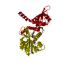
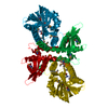
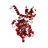
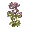
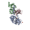
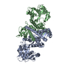
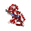
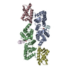
| Title | CRYSTAL STRUCTURE ANALYSIS OF ISOFLAVONE O-METHYLTRANSFERASE | ||||||
 Components Components | ISOFLAVONE O-METHYLTRANSFERASE | ||||||
 Keywords Keywords |  TRANSFERASE / protein-product complex TRANSFERASE / protein-product complex | ||||||
| Function / homology |  Function and homology informationisoflavone 7-O-methyltransferase / isoflavone 7-O-methyltransferase activity / methylation / protein dimerization activity Function and homology informationisoflavone 7-O-methyltransferase / isoflavone 7-O-methyltransferase activity / methylation / protein dimerization activitySimilarity search - Function | ||||||
| Biological species |  Medicago sativa (alfalfa) Medicago sativa (alfalfa) | ||||||
| Method | X-RAY DIFFRACTION / SYNCHROTRON / Resolution: 1.4 Å | ||||||
 Authors Authors | Zubieta, C. / Dixon, R.A. / Noel, J.P. | ||||||
 Citation Citation | Journal: Nat.Struct.Biol. / Year: 2001 Title: Structures of two natural product methyltransferases reveal the basis for substrate specificity in plant O-methyltransferases. Authors: Zubieta, C. / He, X.Z. / Dixon, R.A. / Noel, J.P. | ||||||
| History |
|
- Structure visualization
| Structure viewer | Molecule: MolmilJmol/JSmol |
|---|
- Downloads & links
-Download
| PDBx/mmCIF format | 1fp2.cif.gz | 87.8 KB | Display | PDBx/mmCIF format |
|---|---|---|---|---|
| PDB format | pdb1fp2.ent.gz | 65.1 KB | Display | PDB format |
| PDBx/mmJSON format | 1fp2.json.gz | Tree view | PDBx/mmJSON format | |
| Others |  Other downloads Other downloads |
-Validation report
| Arichive directory | https://data.pdbj.org/pub/pdb/validation_reports/fp/1fp2ftp://data.pdbj.org/pub/pdb/validation_reports/fp/1fp2 | HTTPS FTP |
|---|
-Related structure data


-Links
 PDBj
PDBj- Assembly
| Deposited unit | 
| ||||||||
|---|---|---|---|---|---|---|---|---|---|
| 1 | 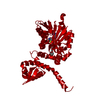
| ||||||||
| Unit cell |
| ||||||||
| Details | The biological assembly is a dimer constructed from chain A by a two-fold rotation. |
-Components
| #1: Protein | Mass: 39648.715 Da / Num. of mol.: 1 Source method: isolated from a genetically manipulated source Source: (gene. exp.) Medicago sativa (alfalfa) / Production host:  Escherichia coli (E. coli) / References: UniProt: O24529 Escherichia coli (E. coli) / References: UniProt: O24529 |
|---|---|
| #2: Chemical | ChemComp-SAH / S-Adenosyl-L-homocysteine  Type: L-peptide linking / Mass: 384.411 Da / Num. of mol.: 1 / Source method: obtained synthetically / Formula: C14H20N6O5S Type: L-peptide linking / Mass: 384.411 Da / Num. of mol.: 1 / Source method: obtained synthetically / Formula: C14H20N6O5S |
| #3: Chemical | ChemComp-HMO /   Mass: 268.264 Da / Num. of mol.: 1 / Source method: obtained synthetically / Formula: C16H12O4 Mass: 268.264 Da / Num. of mol.: 1 / Source method: obtained synthetically / Formula: C16H12O4 |
| #4: Water | ChemComp-HOH / Water Mass: 18.015 Da / Num. of mol.: 247 / Source method: isolated from a natural source / Formula: H2O Mass: 18.015 Da / Num. of mol.: 247 / Source method: isolated from a natural source / Formula: H2O |
-Experimental details
-Experiment
| Experiment | Method: X-RAY DIFFRACTION / Number of used crystals: 1 |
|---|
- Sample preparation
| Crystal | Density Matthews: 2.83 Å3/Da / Density % sol: 56.61 % | |||||||||||||||||||||||||
|---|---|---|---|---|---|---|---|---|---|---|---|---|---|---|---|---|---|---|---|---|---|---|---|---|---|---|
| Crystal grow | Temperature: 277 K / Method: vapor diffusion, hanging drop / pH: 8.25 Details: PEG 8000, lithium sulfate, pH 8.25, VAPOR DIFFUSION, HANGING DROP, temperature 277K | |||||||||||||||||||||||||
| Crystal grow | *PLUS Temperature: 15 ℃ | |||||||||||||||||||||||||
| Components of the solutions | *PLUS
|
-Data collection
| Diffraction | Mean temperature: 105 K |
|---|---|
| Diffraction source | Source: SYNCHROTRON / Site: SSRL  / Beamline: BL9-2 / Wavelength: 0.92 / Beamline: BL9-2 / Wavelength: 0.92 |
| Detector | Type: ADSC QUANTUM 4 / Detector: CCD / Date: Feb 16, 2000 |
| Radiation | Protocol: SINGLE WAVELENGTH / Monochromatic (M) / Laue (L): M / Scattering type: x-ray |
| Radiation wavelength | Wavelength: 0.92 Å / Relative weight: 1 |
| Reflection | Resolution: 1.4→99 Å / Num. all: 87599 / Num. obs: 82735 / % possible obs: 94.4 % / Observed criterion σ(F): 1.5 / Observed criterion σ(I): 1.5 / Redundancy: 1.8 % / Biso Wilson estimate: 17.8 Å2 / Rmerge(I) obs: 0.04 / Net I/σ(I): 19.1 |
| Reflection shell | Resolution: 1.4→1.42 Å / Redundancy: 1.9 % / Rmerge(I) obs: 0.65 / Num. unique all: 6859 / % possible all: 65.5 |
| Reflection | *PLUS Num. measured all: 152679 / Rmerge(I) obs: 0.04 |
| Reflection shell | *PLUS % possible obs: 65 % / Rmerge(I) obs: 0.7 / Mean I/σ(I) obs: 1.3 |
- Processing
| Software |
| ||||||||||||||||||||||||||||||||||||||||
|---|---|---|---|---|---|---|---|---|---|---|---|---|---|---|---|---|---|---|---|---|---|---|---|---|---|---|---|---|---|---|---|---|---|---|---|---|---|---|---|---|---|
| Refinement | Resolution: 1.4→69.71 Å / Rfactor Rfree error: 0.004 / Data cutoff high absF: 1360469.45 / Data cutoff low absF: 0 / Isotropic thermal model: RESTRAINED / Cross valid method: THROUGHOUT / σ(F): 0 / σ(I): 1.5 / Stereochemistry target values: Engh & Huber
| ||||||||||||||||||||||||||||||||||||||||
| Solvent computation | Solvent model: FLAT MODEL / Bsol: 45.34 Å2 / ksol: 0.3714 e/Å3 | ||||||||||||||||||||||||||||||||||||||||
| Displacement parameters | Biso mean: 19.7 Å2
| ||||||||||||||||||||||||||||||||||||||||
| Refine analyze |
| ||||||||||||||||||||||||||||||||||||||||
| Refinement step | Cycle: LAST / Resolution: 1.4→69.71 Å
| ||||||||||||||||||||||||||||||||||||||||
| Refine LS restraints |
| ||||||||||||||||||||||||||||||||||||||||
| LS refinement shell | Resolution: 1.4→1.49 Å / Rfactor Rfree error: 0.017 / Total num. of bins used: 6
| ||||||||||||||||||||||||||||||||||||||||
| Xplor file |
| ||||||||||||||||||||||||||||||||||||||||
| Software | *PLUS Name: CNS / Version: 1 / Classification: refinement | ||||||||||||||||||||||||||||||||||||||||
| Refinement | *PLUS σ(F): 0 / % reflection Rfree: 5 % | ||||||||||||||||||||||||||||||||||||||||
| Solvent computation | *PLUS | ||||||||||||||||||||||||||||||||||||||||
| Displacement parameters | *PLUS Biso mean: 19.7 Å2 | ||||||||||||||||||||||||||||||||||||||||
| Refine LS restraints | *PLUS
| ||||||||||||||||||||||||||||||||||||||||
| LS refinement shell | *PLUS Rfactor Rfree: 0.413 / % reflection Rfree: 4.8 % / Rfactor Rwork: 0.37 |
