 Movie
Movie Controller

[English] 日本語
 Yorodumi
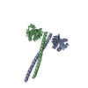
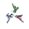
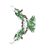
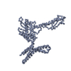
- PDB-5al7: N-terminal fragment of Drosophila melanogaster Sas-6 (F143D), dim... -
+ Open data

- Basic information
| Entry | Database: PDB / ID: 5al7 | ||||||
|---|---|---|---|---|---|---|---|
| Title | N-terminal fragment of Drosophila melanogaster Sas-6 (F143D), dimerised via the coiled-coil domain. | ||||||
 Components Components | SPINDLE ASSEMBLY ABNORMAL PROTEIN 6 HOMOLOG | ||||||
 Keywords Keywords |  STRUCTURAL PROTEIN / CENTRIOLE / CARTWHEEL STRUCTURAL PROTEIN / CENTRIOLE / CARTWHEEL | ||||||
| Function / homology |  Function and homology information Function and homology informationciliary basal body organization / centriole assembly / centriole-centriole cohesion / centrosome cycle / centriole replication / centrosome duplication / centriole / ciliary basal body / centrosome / cytoplasmSimilarity search - Function | ||||||
| Biological species |  DROSOPHILA MELANOGASTER (fruit fly) DROSOPHILA MELANOGASTER (fruit fly) | ||||||
| Method | X-RAY DIFFRACTION / SYNCHROTRON / MOLECULAR REPLACEMENT / Resolution: 2.92 Å | ||||||
 Authors Authors | Cottee, M.A. / Johnson, S. / Lea, S.M. | ||||||
 Citation Citation | Journal: Elife / Year: 2015 Title: The homo-oligomerisation of both Sas-6 and Ana2 is required for efficient centriole assembly in flies. Authors: Cottee, M.A. / Muschalik, N. / Johnson, S. / Leveson, J. / Raff, J.W. / Lea, S.M. | ||||||
| History |
| ||||||
| Remark 700 | SHEET DETERMINATION METHOD: DSSP THE SHEETS PRESENTED AS "AA" IN EACH CHAIN ON SHEET RECORDS BELOW ... SHEET DETERMINATION METHOD: DSSP THE SHEETS PRESENTED AS "AA" IN EACH CHAIN ON SHEET RECORDS BELOW IS ACTUALLY AN 7-STRANDED BARREL THIS IS REPRESENTED BY A 8-STRANDED SHEET IN WHICH THE FIRST AND LAST STRANDS ARE IDENTICAL. THE SHEETS PRESENTED AS "BA" IN EACH CHAIN ON SHEET RECORDS BELOW IS ACTUALLY AN 7-STRANDED BARREL THIS IS REPRESENTED BY A 8-STRANDED SHEET IN WHICH THE FIRST AND LAST STRANDS ARE IDENTICAL. |
- Structure visualization
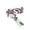
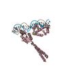
| Structure viewer | Molecule: MolmilJmol/JSmol |
|---|
- Downloads & links
-Download
| PDBx/mmCIF format | 5al7.cif.gz | 182.2 KB | Display | PDBx/mmCIF format |
|---|---|---|---|---|
| PDB format | pdb5al7.ent.gz | 147.5 KB | Display | PDB format |
| PDBx/mmJSON format | 5al7.json.gz | Tree view | PDBx/mmJSON format | |
| Others |  Other downloads Other downloads |
-Validation report
| Arichive directory | https://data.pdbj.org/pub/pdb/validation_reports/al/5al7ftp://data.pdbj.org/pub/pdb/validation_reports/al/5al7 | HTTPS FTP |
|---|
-Related structure data
| Related structure data |  5al6C  2y3vS  2y3wS  3q0xS C: citing same article ( S: Starting model for refinement |
|---|---|
| Similar structure data |
-Links
 PDBj
PDBj- Assembly
| Deposited unit | 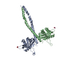
| ||||||||||||
|---|---|---|---|---|---|---|---|---|---|---|---|---|---|
| 1 |
| ||||||||||||
| Unit cell |
| ||||||||||||
| Noncrystallographic symmetry (NCS) | NCS oper:
|
-Components
| #1: Protein | Mass: 25168.453 Da / Num. of mol.: 2 Fragment: N-TERMINAL HEAD GROUP PLUS COILED-COIL, UNP RESIDUES 2-216 Mutation: YES Source method: isolated from a genetically manipulated source Source: (gene. exp.) DROSOPHILA MELANOGASTER (fruit fly) / Plasmid: PETM-14 / Production host:  ESCHERICHIA COLI B (bacteria) / Variant (production host): B834 (DE3) / References: UniProt: Q9VAC8 ESCHERICHIA COLI B (bacteria) / Variant (production host): B834 (DE3) / References: UniProt: Q9VAC8#2: Chemical | Nitrate  Mass: 62.005 Da / Num. of mol.: 2 / Source method: obtained synthetically / Formula: NO3 Mass: 62.005 Da / Num. of mol.: 2 / Source method: obtained synthetically / Formula: NO3#3: Water | ChemComp-HOH / | Water Mass: 18.015 Da / Num. of mol.: 33 / Source method: isolated from a natural source / Formula: H2O Mass: 18.015 Da / Num. of mol.: 33 / Source method: isolated from a natural source / Formula: H2OSequence details | AAL68137 VARIANT, E36D, I207L | |
|---|
-Experimental details
-Experiment
| Experiment | Method: X-RAY DIFFRACTION / Number of used crystals: 1 |
|---|
- Sample preparation
| Crystal | Density Matthews: 3.74 Å3/Da / Density % sol: 67.09 % / Description: NONE |
|---|---|
| Crystal grow | pH: 7.5 Details: 0.1M BICINE/TRIZMA MIX (PH 8.5), 20% W/V PEG 550MME, 10% W/V PEG 20K, 30 MM NANO3, 30 MM NA2HPO4, 30 MM (NH4)2SO4 |
-Data collection
| Diffraction | Mean temperature: 100 K |
|---|---|
| Diffraction source | Source: SYNCHROTRON / Site: ESRF  / Beamline: ID23-2 / Wavelength: 0.8726 / Beamline: ID23-2 / Wavelength: 0.8726 |
| Detector | Date: Sep 10, 2011 |
| Radiation | Protocol: SINGLE WAVELENGTH / Monochromatic (M) / Laue (L): M / Scattering type: x-ray |
| Radiation wavelength | Wavelength: 0.8726 Å / Relative weight: 1 |
| Reflection | Resolution: 2.92→41.43 Å / Num. obs: 15767 / % possible obs: 97.4 % / Observed criterion σ(I): 2 / Redundancy: 5.2 % / Biso Wilson estimate: 43.17 Å2 / Rmerge(I) obs: 0.13 / Net I/σ(I): 12.1 |
| Reflection shell | Resolution: 2.92→3 Å / Redundancy: 5.1 % / Rmerge(I) obs: 0.67 / Mean I/σ(I) obs: 2.6 / % possible all: 96.4 |
- Processing
| Software |
| ||||||||||||||||||||||||||||||||||||||||||||||||||||||||||||||||||||||||||||||||||||||||||||||||||||||||||||||||||||||||||||||||||||||||||||||||||||||||||||||||||||||||||||||||||||||||||||||||||||||||
|---|---|---|---|---|---|---|---|---|---|---|---|---|---|---|---|---|---|---|---|---|---|---|---|---|---|---|---|---|---|---|---|---|---|---|---|---|---|---|---|---|---|---|---|---|---|---|---|---|---|---|---|---|---|---|---|---|---|---|---|---|---|---|---|---|---|---|---|---|---|---|---|---|---|---|---|---|---|---|---|---|---|---|---|---|---|---|---|---|---|---|---|---|---|---|---|---|---|---|---|---|---|---|---|---|---|---|---|---|---|---|---|---|---|---|---|---|---|---|---|---|---|---|---|---|---|---|---|---|---|---|---|---|---|---|---|---|---|---|---|---|---|---|---|---|---|---|---|---|---|---|---|---|---|---|---|---|---|---|---|---|---|---|---|---|---|---|---|---|---|---|---|---|---|---|---|---|---|---|---|---|---|---|---|---|---|---|---|---|---|---|---|---|---|---|---|---|---|---|---|---|---|
| Refinement | Method to determine structure: MOLECULAR REPLACEMENT Starting model: ENSEMBLE OF PDB ENTRIES 2Y3V A,B,D, 2Y3W A, B 3Q0X A,B Resolution: 2.92→41.429 Å / SU ML: 0.31 / σ(F): 1.34 / Phase error: 24.84 / Stereochemistry target values: ML
| ||||||||||||||||||||||||||||||||||||||||||||||||||||||||||||||||||||||||||||||||||||||||||||||||||||||||||||||||||||||||||||||||||||||||||||||||||||||||||||||||||||||||||||||||||||||||||||||||||||||||
| Solvent computation | Shrinkage radii: 0.9 Å / VDW probe radii: 1.11 Å / Solvent model: FLAT BULK SOLVENT MODEL | ||||||||||||||||||||||||||||||||||||||||||||||||||||||||||||||||||||||||||||||||||||||||||||||||||||||||||||||||||||||||||||||||||||||||||||||||||||||||||||||||||||||||||||||||||||||||||||||||||||||||
| Refinement step | Cycle: LAST / Resolution: 2.92→41.429 Å
| ||||||||||||||||||||||||||||||||||||||||||||||||||||||||||||||||||||||||||||||||||||||||||||||||||||||||||||||||||||||||||||||||||||||||||||||||||||||||||||||||||||||||||||||||||||||||||||||||||||||||
| Refine LS restraints |
| ||||||||||||||||||||||||||||||||||||||||||||||||||||||||||||||||||||||||||||||||||||||||||||||||||||||||||||||||||||||||||||||||||||||||||||||||||||||||||||||||||||||||||||||||||||||||||||||||||||||||
| LS refinement shell |
| ||||||||||||||||||||||||||||||||||||||||||||||||||||||||||||||||||||||||||||||||||||||||||||||||||||||||||||||||||||||||||||||||||||||||||||||||||||||||||||||||||||||||||||||||||||||||||||||||||||||||
| Refinement TLS params. | Method: refined / Refine-ID: X-RAY DIFFRACTION
| ||||||||||||||||||||||||||||||||||||||||||||||||||||||||||||||||||||||||||||||||||||||||||||||||||||||||||||||||||||||||||||||||||||||||||||||||||||||||||||||||||||||||||||||||||||||||||||||||||||||||
| Refinement TLS group |
|
