 Movie
Movie Controller


+ Open data

- Basic information
| Entry | Database: PDB / ID: 5a2e | ||||||
|---|---|---|---|---|---|---|---|
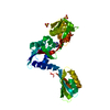
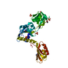
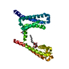
| Title | Extracellular SRCR domains of human CD6 | ||||||
 Components Components | T-CELL DIFFERENTIATION ANTIGEN CD6 | ||||||
 Keywords Keywords |  IMMUNE SYSTEM / SCAVENGER RECEPTOR CYSTEINE RICH / SRCR IMMUNE SYSTEM / SCAVENGER RECEPTOR CYSTEINE RICH / SRCR | ||||||
| Function / homology |  Function and homology information Function and homology information: / lipoteichoic acid binding / acute inflammatory response to antigenic stimulus / immunological synapse formation / scavenger receptor activity / positive regulation of cytokine production involved in inflammatory response / heterophilic cell-cell adhesion via plasma membrane cell adhesion molecules / plasma membrane => GO:0005886 / immunological synapse / positive regulation of T cell proliferation ...: / lipoteichoic acid binding / acute inflammatory response to antigenic stimulus / immunological synapse formation / scavenger receptor activity / positive regulation of cytokine production involved in inflammatory response / heterophilic cell-cell adhesion via plasma membrane cell adhesion molecules / plasma membrane => GO:0005886 / immunological synapse / positive regulation of T cell proliferation / lipopolysaccharide-mediated signaling pathway / lipopolysaccharide binding / adaptive immune response / response to lipopolysaccharide / external side of plasma membrane / innate immune response / extracellular region / identical protein bindingSimilarity search - Function | ||||||
| Biological species |  HOMO SAPIENS (human) HOMO SAPIENS (human) | ||||||
| Method | X-RAY DIFFRACTION / SYNCHROTRON / MOLECULAR REPLACEMENT / Resolution: 3.15 Å | ||||||
 Authors Authors | Chappell, P.E. / Johnson, S. / Lea, S.M. / Brown, M.H. | ||||||
 Citation Citation | Journal: Structure / Year: 2015 Title: Structures of Cd6 and its Ligand Cd166 Give Insight Into Their Interaction. Authors: Chappell, P.E. / Garner, L.I. / Yan, J. / Metcalfe, C. / Hatherley, D. / Johnson, S. / Robinson, C.V. / Lea, S.M. / Brown, M.H. | ||||||
| History |
|
- Structure visualization
| Structure viewer | Molecule: MolmilJmol/JSmol |
|---|
- Downloads & links
-Download
| PDBx/mmCIF format | 5a2e.cif.gz | 72.4 KB | Display | PDBx/mmCIF format |
|---|---|---|---|---|
| PDB format | pdb5a2e.ent.gz | 53.5 KB | Display | PDB format |
| PDBx/mmJSON format | 5a2e.json.gz | Tree view | PDBx/mmJSON format | |
| Others |  Other downloads Other downloads |
-Validation report
| Arichive directory | https://data.pdbj.org/pub/pdb/validation_reports/a2/5a2eftp://data.pdbj.org/pub/pdb/validation_reports/a2/5a2e | HTTPS FTP |
|---|
-Related structure data
| Related structure data | 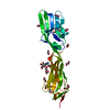 5a2fC  1by2S C: citing same article ( S: Starting model for refinement |
|---|---|
| Similar structure data |


-Links
 PDBj
PDBj- Assembly
| Deposited unit | 
| ||||||||
|---|---|---|---|---|---|---|---|---|---|
| 1 |
| ||||||||
| Unit cell |
|
-Components
| #1: Protein | Mass: 39613.168 Da / Num. of mol.: 1 / Fragment: EXTRACELLULAR SRCR DOMAINS Source method: isolated from a genetically manipulated source Source: (gene. exp.) HOMO SAPIENS (human) / Cell line: CHO LEC 3.2.8.1 / Production host:   CRICETULUS GRISEUS (Chinese hamster) / References: UniProt: P30203 CRICETULUS GRISEUS (Chinese hamster) / References: UniProt: P30203 | ||
|---|---|---|---|
| #2: Sugar | ChemComp-NAG / N-Acetylglucosamine  Type: D-saccharide, beta linking / Mass: 221.208 Da / Num. of mol.: 1 Type: D-saccharide, beta linking / Mass: 221.208 Da / Num. of mol.: 1Source method: isolated from a genetically manipulated source Formula: C8H15NO6 | ||
| #3: Chemical | ChemComp-EDO / Ethylene glycol  Mass: 62.068 Da / Num. of mol.: 6 / Source method: obtained synthetically / Formula: C2H6O2 Mass: 62.068 Da / Num. of mol.: 6 / Source method: obtained synthetically / Formula: C2H6O2#4: Water | ChemComp-HOH / | Water Mass: 18.015 Da / Num. of mol.: 43 / Source method: isolated from a natural source / Formula: H2O Mass: 18.015 Da / Num. of mol.: 43 / Source method: isolated from a natural source / Formula: H2O |
-Experimental details
-Experiment
| Experiment | Method: X-RAY DIFFRACTION / Number of used crystals: 1 |
|---|
- Sample preparation
| Crystal | Density Matthews: 5.46 Å3/Da / Density % sol: 77.49 % / Description: NONE |
|---|---|
| Crystal grow | pH: 6.5 Details: 0.1M AMMONIUM SULFATE, 0.3M SODIUM FORMATE, 0.1M SODIUM CACODYLATE, 3% W/V PGA-LM, 20% MPD, PH 6.5 |
-Data collection
| Diffraction | Mean temperature: 100 K |
|---|---|
| Diffraction source | Source: SYNCHROTRON / Site: Diamond  / Beamline: I04-1 / Wavelength: 0.92 / Beamline: I04-1 / Wavelength: 0.92 |
| Detector | Type: DECTRIS PILATUS / Detector: PIXEL / Date: Jan 31, 2013 |
| Radiation | Protocol: SINGLE WAVELENGTH / Monochromatic (M) / Laue (L): M / Scattering type: x-ray |
| Radiation wavelength | Wavelength: 0.92 Å / Relative weight: 1 |
| Reflection | Resolution: 3.15→77.93 Å / Num. obs: 12884 / % possible obs: 99.6 % / Observed criterion σ(I): 1 / Redundancy: 5.4 % / Biso Wilson estimate: 71.97 Å2 / Rmerge(I) obs: 0.23 / Net I/σ(I): 7.2 |
| Reflection shell | Resolution: 3.15→3.23 Å / Redundancy: 5.5 % / Rmerge(I) obs: 0.8 / Mean I/σ(I) obs: 2.2 / % possible all: 99.8 |
- Processing
| Software |
| ||||||||||||||||||||||||||||||||||||||||||||||||||||||||||||||||||||||||||||||||||||||||||||||||||||||||||||||||||
|---|---|---|---|---|---|---|---|---|---|---|---|---|---|---|---|---|---|---|---|---|---|---|---|---|---|---|---|---|---|---|---|---|---|---|---|---|---|---|---|---|---|---|---|---|---|---|---|---|---|---|---|---|---|---|---|---|---|---|---|---|---|---|---|---|---|---|---|---|---|---|---|---|---|---|---|---|---|---|---|---|---|---|---|---|---|---|---|---|---|---|---|---|---|---|---|---|---|---|---|---|---|---|---|---|---|---|---|---|---|---|---|---|---|---|---|
| Refinement | Method to determine structure: MOLECULAR REPLACEMENT Starting model: PDB ENTRY 1BY2 Resolution: 3.15→77.93 Å / Cor.coef. Fo:Fc: 0.8352 / Cor.coef. Fo:Fc free: 0.7738 / Cross valid method: THROUGHOUT / σ(F): 0 / SU R Blow DPI: 0.715 / SU Rfree Blow DPI: 0.384
| ||||||||||||||||||||||||||||||||||||||||||||||||||||||||||||||||||||||||||||||||||||||||||||||||||||||||||||||||||
| Displacement parameters | Biso mean: 37.69 Å2
| ||||||||||||||||||||||||||||||||||||||||||||||||||||||||||||||||||||||||||||||||||||||||||||||||||||||||||||||||||
| Refine analyze | Luzzati coordinate error obs: 0.731 Å | ||||||||||||||||||||||||||||||||||||||||||||||||||||||||||||||||||||||||||||||||||||||||||||||||||||||||||||||||||
| Refinement step | Cycle: LAST / Resolution: 3.15→77.93 Å
| ||||||||||||||||||||||||||||||||||||||||||||||||||||||||||||||||||||||||||||||||||||||||||||||||||||||||||||||||||
| Refine LS restraints |
| ||||||||||||||||||||||||||||||||||||||||||||||||||||||||||||||||||||||||||||||||||||||||||||||||||||||||||||||||||
| LS refinement shell | Resolution: 3.15→3.45 Å / Total num. of bins used: 6
|
