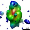
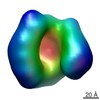
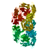
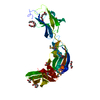
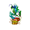
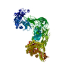
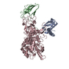
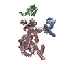
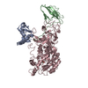
Journal: Proc Natl Acad Sci U S A / Year: 2015 Title: Structural and functional insights into Escherichia coli α2-macroglobulin endopeptidase snap-trap inhibition. Authors: Irene Garcia-Ferrer / Pedro Arêde / Josué Gómez-Blanco / Daniel Luque / Stephane Duquerroy / José R Castón / Theodoros Goulas / F Xavier Gomis-Rüth / Abstract: The survival of commensal bacteria requires them to evade host peptidases. Gram-negative bacteria from the human gut microbiome encode a relative of the human endopeptidase inhibitor, α2- ...The survival of commensal bacteria requires them to evade host peptidases. Gram-negative bacteria from the human gut microbiome encode a relative of the human endopeptidase inhibitor, α2-macroglobulin (α2M). Escherichia coli α2M (ECAM) is a ∼ 180-kDa multidomain membrane-anchored pan-peptidase inhibitor, which is cleaved by host endopeptidases in an accessible bait region. Structural studies by electron microscopy and crystallography reveal that this cleavage causes major structural rearrangement of more than half the 13-domain structure from a native to a compact induced form. It also exposes a reactive thioester bond, which covalently traps the peptidase. Subsequently, peptidase-laden ECAM is shed from the membrane and may dimerize. Trapped peptidases are still active except against very large substrates, so inhibition potentially prevents damage of large cell envelope components, but not host digestion. Mechanistically, these results document a novel monomeric "snap trap."
Resolution: 2.7→48 Å / Num. all: 28350 / Num. obs: 28350 / % possible obs: 99.4 % / Redundancy: 6.4 % / Biso Wilson estimate: 86 Å2 / Rmerge(I) obs: 0.048 / Net I/σ(I): 24.3
Reflection shell
Resolution: 2.7→2.77 Å / Redundancy: 6.6 % / Rmerge(I) obs: 0.654 / Mean I/σ(I) obs: 2.9 / % possible all: 95.3
-
Processing
Software
Name
Version
Classification
XDS
datareduction
XSCALE
datascaling
SHELXDE
phasing
Coot
modelbuilding
BUSTER
2.11.5
refinement
Refinement
Method to determine structure: SIRAS Starting model: dataset of a Se-Met crystal collected at the selenium absorption peak and a native dataset to higher resolution Resolution: 2.7→47.98 Å / Cor.coef. Fo:Fc: 0.9405 / Cor.coef. Fo:Fc free: 0.8812 / SU R Cruickshank DPI: 0.404 / Cross valid method: THROUGHOUT / σ(F): 0 / SU R Blow DPI: 0.398 / SU Rfree Blow DPI: 0.268 / SU Rfree Cruickshank DPI: 0.273
Rfactor
Num. reflection
% reflection
Selection details
Rfree
0.2399
747
2.64 %
RANDOM
Rwork
0.1903
-
-
-
obs
0.1916
28349
99.44 %
-
Displacement parameters
Biso mean: 80.34 Å2
Baniso -1
Baniso -2
Baniso -3
1-
-18.4778 Å2
0 Å2
0 Å2
2-
-
11.2781 Å2
0 Å2
3-
-
-
7.1997 Å2
Refine analyze
Luzzati coordinate error obs: 0.429 Å
Refinement step
Cycle: 1 / Resolution: 2.7→47.98 Å
Protein
Nucleic acid
Ligand
Solvent
Total
Num. atoms
4920
0
32
96
5048
Refine LS restraints
Refine-ID
Type
Dev ideal
Number
Restraint function
Weight
X-RAY DIFFRACTION
t_bond_d
0.01
5044
HARMONIC
2
X-RAY DIFFRACTION
t_angle_deg
1.16
6860
HARMONIC
2
X-RAY DIFFRACTION
t_dihedral_angle_d
2343
SINUSOIDAL
2
X-RAY DIFFRACTION
t_incorr_chiral_ct
X-RAY DIFFRACTION
t_pseud_angle
X-RAY DIFFRACTION
t_trig_c_planes
144
HARMONIC
2
X-RAY DIFFRACTION
t_gen_planes
725
HARMONIC
5
X-RAY DIFFRACTION
t_it
5044
HARMONIC
20
X-RAY DIFFRACTION
t_nbd
0
SEMIHARMONIC
5
X-RAY DIFFRACTION
t_omega_torsion
3.16
X-RAY DIFFRACTION
t_other_torsion
3.28
X-RAY DIFFRACTION
t_improper_torsion
X-RAY DIFFRACTION
t_chiral_improper_torsion
649
SEMIHARMONIC
5
X-RAY DIFFRACTION
t_sum_occupancies
3
HARMONIC
1
X-RAY DIFFRACTION
t_utility_distance
X-RAY DIFFRACTION
t_utility_angle
X-RAY DIFFRACTION
t_utility_torsion
X-RAY DIFFRACTION
t_ideal_dist_contact
5459
SEMIHARMONIC
4
LS refinement shell
Resolution: 2.7→2.8 Å / Total num. of bins used: 14
Rfactor
Num. reflection
% reflection
Rfree
0.3203
74
2.59 %
Rwork
0.2582
2783
-
all
0.2598
2857
-
obs
-
-
99.44 %
Refinement TLS params.
Method: refined / Refine-ID: X-RAY DIFFRACTION
ID
L11 (°2)
L12 (°2)
L13 (°2)
L22 (°2)
L23 (°2)
L33 (°2)
S11 (Å °)
S12 (Å °)
S13 (Å °)
S21 (Å °)
S22 (Å °)
S23 (Å °)
S31 (Å °)
S32 (Å °)
S33 (Å °)
T11 (Å2)
T12 (Å2)
T13 (Å2)
T22 (Å2)
T23 (Å2)
T33 (Å2)
Origin x (Å)
Origin y (Å)
Origin z (Å)
1
4.8501
1.9539
0.7111
1.6623
0.8526
2.5935
-0.1753
0.0739
0.3434
0.1177
0.1648
0.2889
-0.0609
-0.0863
0.0105
-0.0314
-0.0997
-0.0522
-0.0054
-0.0441
0.0309
15.6991
11.024
36.6796
2
3.8716
0.3267
0.0899
6.5698
-0.1432
1.8038
-0.0485
0.3651
-0.0071
-0.3061
0.3028
0.5768
-0.0254
-0.5194
-0.2543
-0.133
-0.0014
-0.1627
0.2274
0.1209
-0.0467
15.5847
54.1342
55.0189
3
1.9666
-0.3603
1.6242
1.6187
-0.4446
4.8131
-0.0713
-0.347
-0.1098
0.0219
0.2278
0.3702
0.0686
-0.8278
-0.1565
-0.1708
0.0126
-0.0005
0.2512
0.0843
-0.0981
21.6294
38.7085
80.7382
4
1.9738
0.7063
0.8498
2.2139
1.4633
3.8366
0.0027
0.2627
-0.4379
0.0157
0.18
-0.2453
0.3763
0.4917
-0.1827
-0.0227
0.0016
-0.0603
0.1463
-0.078
-0.0574
40.1103
24.1796
56.7411
Refinement TLS group
ID
Refine-ID
Refine TLS-ID
Selection details
1
X-RAY DIFFRACTION
1
{A|1015 - 1125 }
2
X-RAY DIFFRACTION
2
{A|1126 - 1170 A|1440 - 1495 }
3
X-RAY DIFFRACTION
3
{A|1171 - 1439 }
4
X-RAY DIFFRACTION
4
{A|1496 - 1653 }
+
About Yorodumi
-
News
-
Feb 9, 2022. New format data for meta-information of EMDB entries
New format data for meta-information of EMDB entries
Version 3 of the EMDB header file is now the official format.
The previous official version 1.9 will be removed from the archive.
In the structure databanks used in Yorodumi, some data are registered as the other names, "COVID-19 virus" and "2019-nCoV". Here are the details of the virus and the list of structure data.
Jan 31, 2019. EMDB accession codes are about to change! (news from PDBe EMDB page)
EMDB accession codes are about to change! (news from PDBe EMDB page)
The allocation of 4 digits for EMDB accession codes will soon come to an end. Whilst these codes will remain in use, new EMDB accession codes will include an additional digit and will expand incrementally as the available range of codes is exhausted. The current 4-digit format prefixed with “EMD-” (i.e. EMD-XXXX) will advance to a 5-digit format (i.e. EMD-XXXXX), and so on. It is currently estimated that the 4-digit codes will be depleted around Spring 2019, at which point the 5-digit format will come into force.
The EM Navigator/Yorodumi systems omit the EMD- prefix.
Related info.:Q: What is EMD? / ID/Accession-code notation in Yorodumi/EM Navigator
Yorodumi is a browser for structure data from EMDB, PDB, SASBDB, etc.
This page is also the successor to EM Navigator detail page, and also detail information page/front-end page for Omokage search.
The word "yorodu" (or yorozu) is an old Japanese word meaning "ten thousand". "mi" (miru) is to see.
Related info.:EMDB / PDB / SASBDB / Comparison of 3 databanks / Yorodumi Search / Aug 31, 2016. New EM Navigator & Yorodumi / Yorodumi Papers / Jmol/JSmol / Function and homology information / Changes in new EM Navigator and Yorodumi
 Movie
Movie Controller

 Yorodumi
 Open data
 Basic information
 Components
 Keywords
 MEMBRANE PROTEIN/INHIBITOR / Bacterial pan-proteinase inhibitor /
  Function and homology information
 Authors
 Spain, 3items
  Citation
 Structure visualization
 Downloads & links
 Other downloads
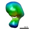


 PDBj
PDBj


 Assembly
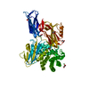

 Mass: 58.693 Da / Num. of mol.: 2 / Source method: obtained synthetically / Formula: Ni
 Mass: 92.094 Da / Num. of mol.: 5 / Source method: obtained synthetically / Formula: C3H8O3
 Mass: 18.015 Da / Num. of mol.: 96 / Source method: isolated from a natural source / Formula: H2O
 Sample preparation
 Processing