 Movie
Movie Controller

[English] 日本語
 Yorodumi
- PDB-4zil: Crystal structure of Rv2466c and oxidoreductase from Mycobacteriu... -
+ Open data

- Basic information
| Entry | Database: PDB / ID: 4zil | ||||||
|---|---|---|---|---|---|---|---|
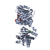
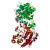
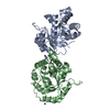
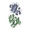
| Title | Crystal structure of Rv2466c and oxidoreductase from Mycobacterium tuberculosis in its reduced state | ||||||
 Components Components | DSBA oxidoreductase | ||||||
 Keywords Keywords |  OXIDOREDUCTASE OXIDOREDUCTASE | ||||||
| Function / homology |  Function and homology information Function and homology information | ||||||
| Biological species |   Mycobacterium tuberculosis (bacteria) Mycobacterium tuberculosis (bacteria) | ||||||
| Method | X-RAY DIFFRACTION / SYNCHROTRON / MOLECULAR REPLACEMENT / Resolution: 1.507 Å | ||||||
 Authors Authors | Albesa-Jove, D. / Urresti, S. / Comino, N. / Tersa, M. / Guerin, M.E. | ||||||
 Citation Citation | Journal: J.Biol.Chem. / Year: 2015 Title: The Redox State Regulates the Conformation of Rv2466c to Activate the Antitubercular Prodrug TP053. Authors: Albesa-Jove, D. / Comino, N. / Tersa, M. / Mohorko, E. / Urresti, S. / Dainese, E. / Chiarelli, L.R. / Pasca, M.R. / Manganelli, R. / Makarov, V. / Riccardi, G. / Svergun, D.I. / Glockshuber, R. / Guerin, M.E. | ||||||
| History |
|
- Structure visualization
| Structure viewer | Molecule: MolmilJmol/JSmol |
|---|
- Downloads & links
-Download
| PDBx/mmCIF format | 4zil.cif.gz | 190.3 KB | Display | PDBx/mmCIF format |
|---|---|---|---|---|
| PDB format | pdb4zil.ent.gz | 150.8 KB | Display | PDB format |
| PDBx/mmJSON format | 4zil.json.gz | Tree view | PDBx/mmJSON format | |
| Others |  Other downloads Other downloads |
-Validation report
| Arichive directory | https://data.pdbj.org/pub/pdb/validation_reports/zi/4zilftp://data.pdbj.org/pub/pdb/validation_reports/zi/4zil | HTTPS FTP |
|---|
-Related structure data
| Related structure data |  4nxiS S: Starting model for refinement |
|---|---|
| Similar structure data |


-Links
 PDBj
PDBj- Assembly
| Deposited unit | 
| ||||||||
|---|---|---|---|---|---|---|---|---|---|
| 1 |
| ||||||||
| Unit cell |
|
-Components
| #1: Protein | Mass: 24589.736 Da / Num. of mol.: 2 Source method: isolated from a genetically manipulated source Source: (gene. exp.) Mycobacterium tuberculosis (H37Rv) (bacteria)Gene: Rv2466c, RVBD_2466c, LH57_13485, P425_02568 / Plasmid: pET23b / Production host: Escherichia coli BL21(DE3) (bacteria) / Variant (production host): pLysS / References: UniProt: O53193#2: Chemical | ChemComp-MG / |   Mass: 24.305 Da / Num. of mol.: 1 / Source method: obtained synthetically / Formula: Mg Mass: 24.305 Da / Num. of mol.: 1 / Source method: obtained synthetically / Formula: Mg#3: Water | ChemComp-HOH / | Water Mass: 18.015 Da / Num. of mol.: 426 / Source method: isolated from a natural source / Formula: H2O Mass: 18.015 Da / Num. of mol.: 426 / Source method: isolated from a natural source / Formula: H2O |
|---|
-Experimental details
-Experiment
| Experiment | Method: X-RAY DIFFRACTION |
|---|
- Sample preparation
| Crystal | Density Matthews: 2 Å3/Da / Density % sol: 38.46 % |
|---|---|
| Crystal grow | Temperature: 291 K / Method: vapor diffusion, sitting drop / pH: 8.5 Details: 0.1 M Tris-HCl pH 8.5, 0.1 M Magnesium chloride, and 17% (w/v) PEG 20,000 |
-Data collection
| Diffraction | Mean temperature: 100 K |
|---|---|
| Diffraction source | Source: SYNCHROTRON / Site: Diamond  / Beamline: I04 / Wavelength: 0.9537 Å / Beamline: I04 / Wavelength: 0.9537 Å |
| Detector | Type: ADSC QUANTUM 315r / Detector: CCD / Date: Oct 20, 2012 |
| Radiation | Monochromator: Double crystal Si(111) / Protocol: SINGLE WAVELENGTH / Monochromatic (M) / Laue (L): M / Scattering type: x-ray |
| Radiation wavelength | Wavelength: 0.9537 Å / Relative weight: 1 |
| Reflection | Resolution: 1.507→40.26 Å / Num. all: 60530 / Num. obs: 60530 / % possible obs: 98.29 % / Observed criterion σ(I): -3 / Redundancy: 4.1 % / Biso Wilson estimate: 11.86 Å2 / Rmerge(I) obs: 0.09497 / Net I/σ(I): 10.47 |
| Reflection shell | Resolution: 1.507→1.561 Å / Redundancy: 3.7 % / Rmerge(I) obs: 0.6503 / Mean I/σ(I) obs: 2.11 / % possible all: 85.07 |
- Processing
| Software |
| |||||||||||||||||||||||||||||||||||||||||||||||||||||||||||||||||||||||||||||||||||||||||||||||||||||||||||||||||||||||||||||||||||||||||||||||||||||||||||||||||
|---|---|---|---|---|---|---|---|---|---|---|---|---|---|---|---|---|---|---|---|---|---|---|---|---|---|---|---|---|---|---|---|---|---|---|---|---|---|---|---|---|---|---|---|---|---|---|---|---|---|---|---|---|---|---|---|---|---|---|---|---|---|---|---|---|---|---|---|---|---|---|---|---|---|---|---|---|---|---|---|---|---|---|---|---|---|---|---|---|---|---|---|---|---|---|---|---|---|---|---|---|---|---|---|---|---|---|---|---|---|---|---|---|---|---|---|---|---|---|---|---|---|---|---|---|---|---|---|---|---|---|---|---|---|---|---|---|---|---|---|---|---|---|---|---|---|---|---|---|---|---|---|---|---|---|---|---|---|---|---|---|---|---|
| Refinement | Method to determine structure: MOLECULAR REPLACEMENT Starting model: 4NXI Resolution: 1.507→40.256 Å / SU ML: 0.12 / Cross valid method: FREE R-VALUE / σ(F): 2 / Phase error: 15.94 / Stereochemistry target values: ML
| |||||||||||||||||||||||||||||||||||||||||||||||||||||||||||||||||||||||||||||||||||||||||||||||||||||||||||||||||||||||||||||||||||||||||||||||||||||||||||||||||
| Solvent computation | Shrinkage radii: 0.9 Å / VDW probe radii: 1.11 Å / Solvent model: FLAT BULK SOLVENT MODEL | |||||||||||||||||||||||||||||||||||||||||||||||||||||||||||||||||||||||||||||||||||||||||||||||||||||||||||||||||||||||||||||||||||||||||||||||||||||||||||||||||
| Displacement parameters | Biso mean: 19.4 Å2 | |||||||||||||||||||||||||||||||||||||||||||||||||||||||||||||||||||||||||||||||||||||||||||||||||||||||||||||||||||||||||||||||||||||||||||||||||||||||||||||||||
| Refinement step | Cycle: LAST / Resolution: 1.507→40.256 Å
| |||||||||||||||||||||||||||||||||||||||||||||||||||||||||||||||||||||||||||||||||||||||||||||||||||||||||||||||||||||||||||||||||||||||||||||||||||||||||||||||||
| Refine LS restraints |
| |||||||||||||||||||||||||||||||||||||||||||||||||||||||||||||||||||||||||||||||||||||||||||||||||||||||||||||||||||||||||||||||||||||||||||||||||||||||||||||||||
| LS refinement shell |
|
