 Movie
Movie Controller


+ Open data

- Basic information
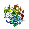
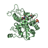
| Entry | Database: PDB / ID: 3hrh | ||||||
|---|---|---|---|---|---|---|---|
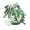
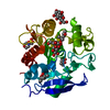
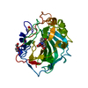
| Title | Crystal Structure of Antigen 85C and Glycerol | ||||||
 Components Components | Antigen 85-C | ||||||
 Keywords Keywords |  HYDROLASE / alpha/beta hydrolase / Acyltransferase / Secreted HYDROLASE / alpha/beta hydrolase / Acyltransferase / Secreted | ||||||
| Function / homology |  Function and homology informationtrehalose O-mycolyltransferase / trehalose O-mycolyltransferase activity / diacylglycerol O-acyltransferase / diacylglycerol O-acyltransferase activity / mycolate cell wall layer assembly / glycolipid biosynthetic process / zymogen binding / lipid transport / acyltransferase activity, transferring groups other than amino-acyl groups / peptidoglycan-based cell wall ...trehalose O-mycolyltransferase / trehalose O-mycolyltransferase activity / diacylglycerol O-acyltransferase / diacylglycerol O-acyltransferase activity / mycolate cell wall layer assembly / glycolipid biosynthetic process / zymogen binding / lipid transport / acyltransferase activity, transferring groups other than amino-acyl groups / peptidoglycan-based cell wall / response to antibiotic / extracellular region Function and homology informationtrehalose O-mycolyltransferase / trehalose O-mycolyltransferase activity / diacylglycerol O-acyltransferase / diacylglycerol O-acyltransferase activity / mycolate cell wall layer assembly / glycolipid biosynthetic process / zymogen binding / lipid transport / acyltransferase activity, transferring groups other than amino-acyl groups / peptidoglycan-based cell wall ...trehalose O-mycolyltransferase / trehalose O-mycolyltransferase activity / diacylglycerol O-acyltransferase / diacylglycerol O-acyltransferase activity / mycolate cell wall layer assembly / glycolipid biosynthetic process / zymogen binding / lipid transport / acyltransferase activity, transferring groups other than amino-acyl groups / peptidoglycan-based cell wall / response to antibiotic / extracellular regionSimilarity search - Function | ||||||
| Biological species |   Mycobacterium tuberculosis (bacteria) Mycobacterium tuberculosis (bacteria) | ||||||
| Method | X-RAY DIFFRACTION / SYNCHROTRON / FOURIER SYNTHESIS / Resolution: 2.3 Å | ||||||
 Authors Authors | Boucau, J. / Sanki, A.K. / Umesiri, F.E. / Sucheck, S.J. / Ronning, D.R. | ||||||
 Citation Citation | Journal: Mol Biosyst / Year: 2009 Title: Design, synthesis and biological evaluation of sugar-derived esters, alpha-ketoesters and alpha-ketoamides as inhibitors for Mycobacterium tuberculosis antigen 85C. Authors: Sanki, A.K. / Boucau, J. / Umesiri, F.E. / Ronning, D.R. / Sucheck, S.J. | ||||||
| History |
|
- Structure visualization
| Structure viewer | Molecule: MolmilJmol/JSmol |
|---|
- Downloads & links
-Download
| PDBx/mmCIF format | 3hrh.cif.gz | 123.9 KB | Display | PDBx/mmCIF format |
|---|---|---|---|---|
| PDB format | pdb3hrh.ent.gz | 95.7 KB | Display | PDB format |
| PDBx/mmJSON format | 3hrh.json.gz | Tree view | PDBx/mmJSON format | |
| Others |  Other downloads Other downloads |
-Validation report
| Arichive directory | https://data.pdbj.org/pub/pdb/validation_reports/hr/3hrhftp://data.pdbj.org/pub/pdb/validation_reports/hr/3hrh | HTTPS FTP |
|---|
-Related structure data
| Related structure data | 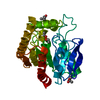 1dqyS S: Starting model for refinement |
|---|---|
| Similar structure data |


-Links
 PDBj
PDBj- Assembly
| Deposited unit | 
| ||||||||
|---|---|---|---|---|---|---|---|---|---|
| 1 | 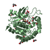
| ||||||||
| 2 | 
| ||||||||
| 3 |
| ||||||||
| Unit cell |
|
-Components
| #1: Protein | Mass: 33242.785 Da / Num. of mol.: 2 Source method: isolated from a genetically manipulated source Source: (gene. exp.) Mycobacterium tuberculosis (bacteria) / Strain: H37Rv / Gene: fbpC, fbpC2, mpt45, MT0137, MTCI5.03c, Rv0129c / Plasmid: pET28 / Production host: Escherichia coli (E. coli) / Strain (production host): BL21References: UniProt: P0A4V4, UniProt: P9WQN9*PLUS, Transferases; Acyltransferases; Transferring groups other than aminoacyl groups#2: Chemical | ChemComp-GOL / Glycerol  Mass: 92.094 Da / Num. of mol.: 20 / Source method: obtained synthetically / Formula: C3H8O3 Mass: 92.094 Da / Num. of mol.: 20 / Source method: obtained synthetically / Formula: C3H8O3#3: Water | ChemComp-HOH / | Water Mass: 18.015 Da / Num. of mol.: 103 / Source method: isolated from a natural source / Formula: H2O Mass: 18.015 Da / Num. of mol.: 103 / Source method: isolated from a natural source / Formula: H2O |
|---|
-Experimental details
-Experiment
| Experiment | Method: X-RAY DIFFRACTION / Number of used crystals: 1 |
|---|
- Sample preparation
| Crystal | Density Matthews: 3.05 Å3/Da / Density % sol: 59.64 % |
|---|---|
| Crystal grow | Temperature: 291 K / Method: vapor diffusion, hanging drop / pH: 4.6 Details: 0.1 M Na Acetate and 15% PEG 4000, pH 4.6, VAPOR DIFFUSION, HANGING DROP, temperature 291K |
-Data collection
| Diffraction | Mean temperature: 100 K |
|---|---|
| Diffraction source | Source: SYNCHROTRON / Site: APS  / Beamline: 23-ID-D / Wavelength: 1 Å / Beamline: 23-ID-D / Wavelength: 1 Å |
| Detector | Type: MARMOSAIC 300 mm CCD / Detector: CCD / Date: Jul 11, 2008 |
| Radiation | Protocol: SINGLE WAVELENGTH / Monochromatic (M) / Laue (L): M / Scattering type: x-ray |
| Radiation wavelength | Wavelength: 1 Å / Relative weight: 1 |
| Reflection | Resolution: 2.2→48.44 Å / % possible obs: 99.7 % / Redundancy: 3.7 % / Biso Wilson estimate: 18.5 Å2 / Rsym value: 0.138 / Net I/σ(I): 7.6 |
- Processing
| Software |
| ||||||||||||||||||||||||||||||||||||||||||||||||||||||||||||||||||||||||||||||||
|---|---|---|---|---|---|---|---|---|---|---|---|---|---|---|---|---|---|---|---|---|---|---|---|---|---|---|---|---|---|---|---|---|---|---|---|---|---|---|---|---|---|---|---|---|---|---|---|---|---|---|---|---|---|---|---|---|---|---|---|---|---|---|---|---|---|---|---|---|---|---|---|---|---|---|---|---|---|---|---|---|---|
| Refinement | Method to determine structure: FOURIER SYNTHESIS Starting model: 1DQY Resolution: 2.3→48.44 Å / Rfactor Rfree error: 0.005 / Data cutoff high absF: 85706.77 / Data cutoff low absF: 0 / Isotropic thermal model: RESTRAINED / Cross valid method: THROUGHOUT / σ(F): 0 / Stereochemistry target values: Engh & Huber / Details: BULK SOLVENT MODEL USED
| ||||||||||||||||||||||||||||||||||||||||||||||||||||||||||||||||||||||||||||||||
| Solvent computation | Solvent model: FLAT MODEL / Bsol: 32.4426 Å2 / ksol: 0.4 e/Å3 | ||||||||||||||||||||||||||||||||||||||||||||||||||||||||||||||||||||||||||||||||
| Displacement parameters | Biso mean: 27.3 Å2
| ||||||||||||||||||||||||||||||||||||||||||||||||||||||||||||||||||||||||||||||||
| Refine analyze |
| ||||||||||||||||||||||||||||||||||||||||||||||||||||||||||||||||||||||||||||||||
| Refinement step | Cycle: LAST / Resolution: 2.3→48.44 Å
| ||||||||||||||||||||||||||||||||||||||||||||||||||||||||||||||||||||||||||||||||
| Refine LS restraints |
| ||||||||||||||||||||||||||||||||||||||||||||||||||||||||||||||||||||||||||||||||
| LS refinement shell | Resolution: 2.3→2.44 Å / Rfactor Rfree error: 0.017 / Total num. of bins used: 6
| ||||||||||||||||||||||||||||||||||||||||||||||||||||||||||||||||||||||||||||||||
| Xplor file |
|
