Journal: J Biol Chem / Year: 2015 Title: Structural Basis for Antigen Recognition by Transglutaminase 2-specific Autoantibodies in Celiac Disease. Authors: Xi Chen / Kathrin Hnida / Melissa Ann Graewert / Jan Terje Andersen / Rasmus Iversen / Anne Tuukkanen / Dmitri Svergun / Ludvig M Sollid / Abstract: Antibodies to the autoantigen transglutaminase 2 (TG2) are a hallmark of celiac disease. We have studied the interaction between TG2 and an anti-TG2 antibody (679-14-E06) derived from a single gut ...Antibodies to the autoantigen transglutaminase 2 (TG2) are a hallmark of celiac disease. We have studied the interaction between TG2 and an anti-TG2 antibody (679-14-E06) derived from a single gut IgA plasma cell of a celiac disease patient. The antibody recognizes one of four identified epitopes targeted by antibodies of plasma cells of the disease lesion. The binding interface was identified by small angle x-ray scattering, ab initio and rigid body modeling using the known crystal structure of TG2 and the crystal structure of the antibody Fab fragment, which was solved at 2.4 Å resolution. The result was confirmed by testing binding of the antibody to TG2 mutants by ELISA and surface plasmon resonance. TG2 residues Arg-116 and His-134 were identified to be critical for binding of 679-14-E06 as well as other epitope 1 antibodies. In contrast, antibodies directed toward the two other main epitopes (epitopes 2 and 3) were not affected by these mutations. Molecular dynamics simulations suggest interactions of 679-14-E06 with the N-terminal domain of TG2 via the CDR2 and CDR3 loops of the heavy chain and the CDR2 loop of the light chain. In addition there were contacts of the framework 3 region of the heavy chain with the catalytic domain of TG2. The results provide an explanation for the biased usage of certain heavy and light chain gene segments by epitope 1-specific antibodies in celiac disease.
Mass: 24026.963 Da / Num. of mol.: 1 Source method: isolated from a genetically manipulated source Source: (gene. exp.) Homo sapiens (human) / Production host: Homo sapiens (human)
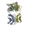
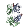
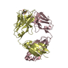
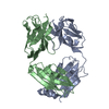
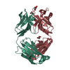
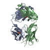
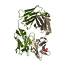
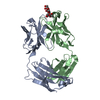
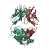
#2: Antibody
679-14-14E06Fabfragmentlightchain
Mass: 23674.209 Da / Num. of mol.: 1 Source method: isolated from a genetically manipulated source Source: (gene. exp.) Homo sapiens (human) / Production host: Homo sapiens (human)
Mass: 18.015 Da / Num. of mol.: 78 / Source method: isolated from a natural source / Formula: H2O
-
Experimental details
-
Experiment
Experiment
Method: X-RAY DIFFRACTION
-
Sample preparation
Crystal
Density Matthews: 2.52 Å3/Da / Density % sol: 51.17 % / Description: crystals look like plates
Crystal grow
Temperature: 293 K / Method: vapor diffusion, hanging drop / pH: 8.5 Details: 0.01 M nickel (II) chloride hexahydrate, 0.1 M Tris pH=8.5, 20% w/v polyethylene glycol monomethyl ether 2000
Protocol: SINGLE WAVELENGTH / Monochromatic (M) / Laue (L): M / Scattering type: x-ray
Radiation wavelength
Wavelength: 0.979093 Å / Relative weight: 1
Reflection
Resolution: 2.4→49.53 Å / Num. obs: 18701 / % possible obs: 99.2 % / Redundancy: 3.4 % / Rsym value: 0.117 / Net I/σ(I): 9.3
Reflection shell
Resolution: 2.4→2.5 Å / Redundancy: 3.5 % / Rmerge(I) obs: 0.605 / Mean I/σ(I) obs: 2.4 / % possible all: 99.5
-
Processing
Software
Name
Version
Classification
PHENIX
1.8.4_1496
refinement
MOSFLM
7.1.0
datareduction
Aimless
5.8.0049
datascaling
PHASER
5.8.0049
phasing
Refinement
Method to determine structure: MOLECULAR REPLACEMENT Starting model: Contant and variable domains of heavy chain PDB code 4HPO and a light chain PDB code 1DFB
In the structure databanks used in Yorodumi, some data are registered as the other names, "COVID-19 virus" and "2019-nCoV". Here are the details of the virus and the list of structure data.
Jan 31, 2019. EMDB accession codes are about to change! (news from PDBe EMDB page)
EMDB accession codes are about to change! (news from PDBe EMDB page)
The allocation of 4 digits for EMDB accession codes will soon come to an end. Whilst these codes will remain in use, new EMDB accession codes will include an additional digit and will expand incrementally as the available range of codes is exhausted. The current 4-digit format prefixed with “EMD-” (i.e. EMD-XXXX) will advance to a 5-digit format (i.e. EMD-XXXXX), and so on. It is currently estimated that the 4-digit codes will be depleted around Spring 2019, at which point the 5-digit format will come into force.
The EM Navigator/Yorodumi systems omit the EMD- prefix.
Related info.:Q: What is EMD? / ID/Accession-code notation in Yorodumi/EM Navigator
Yorodumi is a browser for structure data from EMDB, PDB, SASBDB, etc.
This page is also the successor to EM Navigator detail page, and also detail information page/front-end page for Omokage search.
The word "yorodu" (or yorozu) is an old Japanese word meaning "ten thousand". "mi" (miru) is to see.
Related info.:EMDB / PDB / SASBDB / Comparison of 3 databanks / Yorodumi Search / Aug 31, 2016. New EM Navigator & Yorodumi / Yorodumi Papers / Jmol/JSmol / Function and homology information / Changes in new EM Navigator and Yorodumi
 Movie
Movie Controller

 Yorodumi
 Open data
 Basic information
 Components
 Keywords
 IMMUNE SYSTEM /
  Function and homology information
 Authors
 Norway, 4items
  Citation
 Structure visualization
 Downloads & links
 Other downloads
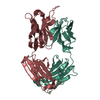


 PDBj
PDBj


 Assembly
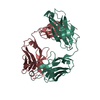
 Mass: 18.015 Da / Num. of mol.: 78 / Source method: isolated from a natural source / Formula: H2O
 Sample preparation
 / Beamline: BM30A / Wavelength: 0.979093 Å
 Processing