 Movie
Movie Controller

[English] 日本語
 Yorodumi
- PDB-4z19: Crystal structure of beta-ketoacyl-ACP synthase III (FabH) from Y... -
+ Open data

- Basic information
| Entry | Database: PDB / ID: 4z19 | ||||||
|---|---|---|---|---|---|---|---|
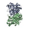
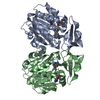
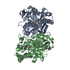
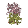
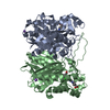
| Title | Crystal structure of beta-ketoacyl-ACP synthase III (FabH) from Yersinia pestis with acetylated active site cysteine | ||||||
 Components Components | 3-oxoacyl-[acyl-carrier-protein] synthase 3 | ||||||
 Keywords Keywords |  TRANSFERASE / FABH / FATTY ACID BIOSYNTHESIS / CONDENSING ENZYME / THIOLASE FOLD / ACETYLATED TRANSFERASE / FABH / FATTY ACID BIOSYNTHESIS / CONDENSING ENZYME / THIOLASE FOLD / ACETYLATED | ||||||
| Function / homology |  Function and homology information Function and homology informationbeta-ketoacyl-[acyl-carrier-protein] synthase III / beta-ketoacyl-acyl-carrier-protein synthase III activity / 3-oxoacyl-[acyl-carrier-protein] synthase activity / fatty acid biosynthetic process / cytoplasmSimilarity search - Function | ||||||
| Biological species |   Yersinia pestis (bacteria) Yersinia pestis (bacteria) | ||||||
| Method | X-RAY DIFFRACTION / SYNCHROTRON / MOLECULAR REPLACEMENT / Resolution: 1.8 Å | ||||||
 Authors Authors | Nanson, J.D. / Forwood, J.K. | ||||||
 Citation Citation | Journal: Sci Rep / Year: 2015 Title: Structural Characterisation of the Beta-Ketoacyl-Acyl Carrier Protein Synthases, FabF and FabH, of Yersinia pestis. Authors: Nanson, J.D. / Himiari, Z. / Swarbrick, C.M. / Forwood, J.K. | ||||||
| History |
|
- Structure visualization
| Structure viewer | Molecule: MolmilJmol/JSmol |
|---|
- Downloads & links
-Download
| PDBx/mmCIF format | 4z19.cif.gz | 133.3 KB | Display | PDBx/mmCIF format |
|---|---|---|---|---|
| PDB format | pdb4z19.ent.gz | 109.2 KB | Display | PDB format |
| PDBx/mmJSON format | 4z19.json.gz | Tree view | PDBx/mmJSON format | |
| Others |  Other downloads Other downloads |
-Validation report
| Arichive directory | https://data.pdbj.org/pub/pdb/validation_reports/z1/4z19ftp://data.pdbj.org/pub/pdb/validation_reports/z1/4z19 | HTTPS FTP |
|---|
-Related structure data


-Links
 PDBj
PDBj

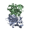
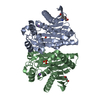
- Assembly
| Deposited unit | 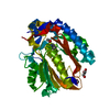
| ||||||||
|---|---|---|---|---|---|---|---|---|---|
| 1 | 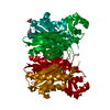
| ||||||||
| Unit cell |
| ||||||||
| Components on special symmetry positions |
|
-Components
| #1: Protein | Mass: 33843.379 Da / Num. of mol.: 1 / Mutation: A229V Source method: isolated from a genetically manipulated source Source: (gene. exp.) Yersinia pestis (bacteria) / Gene: fabH, YPO1597, y1756, YP_2257 / Plasmid: pMCSG21 / Production host: Escherichia coli (E. coli) / Strain (production host): BL21(DE3)PlysSReferences: UniProt: Q8ZFT7, beta-ketoacyl-[acyl-carrier-protein] synthase III | ||
|---|---|---|---|
| #2: Chemical | Glycerol  Mass: 92.094 Da / Num. of mol.: 2 / Source method: obtained synthetically / Formula: C3H8O3 Mass: 92.094 Da / Num. of mol.: 2 / Source method: obtained synthetically / Formula: C3H8O3#3: Water | ChemComp-HOH / | Water Mass: 18.015 Da / Num. of mol.: 230 / Source method: isolated from a natural source / Formula: H2O Mass: 18.015 Da / Num. of mol.: 230 / Source method: isolated from a natural source / Formula: H2O |
-Experimental details
-Experiment
| Experiment | Method: X-RAY DIFFRACTION / Number of used crystals: 1 |
|---|
- Sample preparation
| Crystal | Density Matthews: 1.99 Å3/Da / Density % sol: 38.34 % |
|---|---|
| Crystal grow | Temperature: 296.15 K / Method: vapor diffusion, hanging drop / pH: 7.5 Details: Propan-2-ol, 0.1 M HEPES sodium salt, 15% Glycerol, 24% PEG4000, 2.25mM Acetyl-CoA, 2.25mM Malonyl-CoA |
-Data collection
| Diffraction | Mean temperature: 100 K |
|---|---|
| Diffraction source | Source: SYNCHROTRON / Site: Australian Synchrotron  / Beamline: MX2 / Wavelength: 0.9537 Å / Beamline: MX2 / Wavelength: 0.9537 Å |
| Detector | Type: ADSC QUANTUM 315r / Detector: CCD / Date: Jul 13, 2013 |
| Radiation | Protocol: SINGLE WAVELENGTH / Monochromatic (M) / Laue (L): M / Scattering type: x-ray |
| Radiation wavelength | Wavelength: 0.9537 Å / Relative weight: 1 |
| Reflection | Resolution: 1.8→49.26 Å / Num. obs: 25320 / % possible obs: 99.3 % / Redundancy: 7.2 % / Net I/σ(I): 12.2 |
| Reflection shell | Resolution: 1.8→1.84 Å / Redundancy: 6.6 % / Mean I/σ(I) obs: 4.9 / % possible all: 96.7 |
- Processing
| Software |
| ||||||||||||||||||||||||||||||||||||||||||||||||||||||||||||||||||||||
|---|---|---|---|---|---|---|---|---|---|---|---|---|---|---|---|---|---|---|---|---|---|---|---|---|---|---|---|---|---|---|---|---|---|---|---|---|---|---|---|---|---|---|---|---|---|---|---|---|---|---|---|---|---|---|---|---|---|---|---|---|---|---|---|---|---|---|---|---|---|---|---|
| Refinement | Method to determine structure: MOLECULAR REPLACEMENT / Resolution: 1.8→38.057 Å / SU ML: 0.14 / Cross valid method: FREE R-VALUE / σ(F): 1.34 / Phase error: 15.91 / Stereochemistry target values: ML
| ||||||||||||||||||||||||||||||||||||||||||||||||||||||||||||||||||||||
| Solvent computation | Shrinkage radii: 0.9 Å / VDW probe radii: 1.11 Å / Solvent model: FLAT BULK SOLVENT MODEL | ||||||||||||||||||||||||||||||||||||||||||||||||||||||||||||||||||||||
| Refinement step | Cycle: LAST / Resolution: 1.8→38.057 Å
| ||||||||||||||||||||||||||||||||||||||||||||||||||||||||||||||||||||||
| Refine LS restraints |
| ||||||||||||||||||||||||||||||||||||||||||||||||||||||||||||||||||||||
| LS refinement shell |
| ||||||||||||||||||||||||||||||||||||||||||||||||||||||||||||||||||||||
| Refinement TLS params. | Method: refined / Origin x: 21.2286 Å / Origin y: -15.5349 Å / Origin z: 25.2259 Å
| ||||||||||||||||||||||||||||||||||||||||||||||||||||||||||||||||||||||
| Refinement TLS group | Selection details: all |
