 Movie
Movie Controller

[English] 日本語
 Yorodumi
- PDB-4yzy: Crystal structures reveal transient PERK luminal domain tetrameri... -
+ Open data

- Basic information
| Entry | Database: PDB / ID: 4yzy | |||||||||
|---|---|---|---|---|---|---|---|---|---|---|
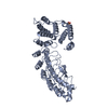
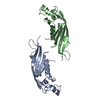
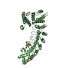
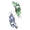
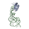
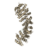
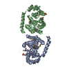
| Title | Crystal structures reveal transient PERK luminal domain tetramerization in ER stress signaling | |||||||||
 Components Components | Eukaryotic translation initiation factor 2-alpha kinase 3 | |||||||||
 Keywords Keywords |  SIGNALING PROTEIN / UPR / ER stress sensing / proteostasis / PERK / dimer SIGNALING PROTEIN / UPR / ER stress sensing / proteostasis / PERK / dimer | |||||||||
| Function / homology |  Function and homology information Function and homology informationPERK regulates gene expression / negative regulation of translation in response to endoplasmic reticulum stress / regulation of endoplasmic reticulum stress-induced eIF2 alpha phosphorylation / positive regulation of glutathione biosynthetic process / negative regulation of translation in response to stress / regulation of endoplasmic reticulum stress-induced intrinsic apoptotic signaling pathway / regulation of translational initiation by eIF2 alpha phosphorylation / eiF2alpha phosphorylation in response to endoplasmic reticulum stress / eukaryotic initiation factor eIF2 binding / chondrocyte development ...PERK regulates gene expression / negative regulation of translation in response to endoplasmic reticulum stress / regulation of endoplasmic reticulum stress-induced eIF2 alpha phosphorylation / positive regulation of glutathione biosynthetic process / negative regulation of translation in response to stress / regulation of endoplasmic reticulum stress-induced intrinsic apoptotic signaling pathway / regulation of translational initiation by eIF2 alpha phosphorylation / eiF2alpha phosphorylation in response to endoplasmic reticulum stress / eukaryotic initiation factor eIF2 binding / chondrocyte development / eukaryotic translation initiation factor 2alpha kinase activity / positive regulation of signal transduction / endoplasmic reticulum quality control compartment / PERK-mediated unfolded protein response / positive regulation of ERAD pathway / SREBP signaling pathway / negative regulation of myelination / positive regulation of endoplasmic reticulum stress-induced intrinsic apoptotic signaling pathway / regulation of fatty acid metabolic process / endocrine pancreas development / endoplasmic reticulum organization / pancreas development / regulation of translational initiation / cellular response to cold / ER overload response / positive regulation of transcription by RNA polymerase I / fat cell differentiation / : / bone mineralization / intrinsic apoptotic signaling pathway in response to endoplasmic reticulum stress / positive regulation of vascular endothelial growth factor production / cellular response to glucose starvation / endoplasmic reticulum unfolded protein response / lactation / cellular response to amino acid starvation / response to endoplasmic reticulum stress / ossification / insulin-like growth factor receptor signaling pathway / skeletal system development / calcium-mediated signaling / Hsp90 protein binding / positive regulation of protein localization to nucleus / peptidyl-serine phosphorylation / protein phosphatase binding / angiogenesis / protein autophosphorylation / negative regulation of translation / non-specific serine/threonine protein kinase / protein kinase activity / translation / protein phosphorylation / negative regulation of gene expression / protein serine kinase activity / protein serine/threonine kinase activity / endoplasmic reticulum membrane / positive regulation of gene expression / negative regulation of apoptotic process / perinuclear region of cytoplasm / enzyme binding / endoplasmic reticulum / ATP binding / identical protein binding / nucleus / cytoplasmSimilarity search - Function | |||||||||
| Biological species |  Mus musculus (house mouse) Mus musculus (house mouse) | |||||||||
| Method | X-RAY DIFFRACTION / SYNCHROTRON / MOLECULAR REPLACEMENT / Resolution: 3.2 Å | |||||||||
 Authors Authors | Carrara, M. / Prischi, F. / Ali, M.M.U. | |||||||||
| Funding support |  United Kingdom, 2items United Kingdom, 2items
| |||||||||
 Citation Citation | Journal: Embo J. / Year: 2015 Title: Crystal structures reveal transient PERK luminal domain tetramerization in endoplasmic reticulum stress signaling. Authors: Carrara, M. / Prischi, F. / Nowak, P.R. / Ali, M.M. | |||||||||
| History |
|
- Structure visualization
| Structure viewer | Molecule: MolmilJmol/JSmol |
|---|
- Downloads & links
-Download
| PDBx/mmCIF format | 4yzy.cif.gz | 79.6 KB | Display | PDBx/mmCIF format |
|---|---|---|---|---|
| PDB format | pdb4yzy.ent.gz | 58.1 KB | Display | PDB format |
| PDBx/mmJSON format | 4yzy.json.gz | Tree view | PDBx/mmJSON format | |
| Others |  Other downloads Other downloads |
-Validation report
| Arichive directory | https://data.pdbj.org/pub/pdb/validation_reports/yz/4yzyftp://data.pdbj.org/pub/pdb/validation_reports/yz/4yzy | HTTPS FTP |
|---|
-Related structure data
| Related structure data |  4yzsSC S: Starting model for refinement C: citing same article ( |
|---|---|
| Similar structure data |


-Links
 PDBj
PDBj- Assembly
| Deposited unit | 
| ||||||||
|---|---|---|---|---|---|---|---|---|---|
| 1 | 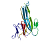
| ||||||||
| Unit cell |
|
-Components
| #1: Protein | Mass: 33509.504 Da / Num. of mol.: 1 / Fragment: UNP residues 100-399 Source method: isolated from a genetically manipulated source Source: (gene. exp.) Mus musculus (house mouse) / Gene: Eif2ak3, Pek, Perk / Production host:  Escherichia coli (E. coli) Escherichia coli (E. coli)References: UniProt: Q9Z2B5, non-specific serine/threonine protein kinase |
|---|
-Experimental details
-Experiment
| Experiment | Method: X-RAY DIFFRACTION |
|---|
- Sample preparation
| Crystal | Density Matthews: 2.67 Å3/Da / Density % sol: 53.92 % |
|---|---|
| Crystal grow | Temperature: 291 K / Method: vapor diffusion, hanging drop Details: 0.1 M MES/imidazole (pH 6.5), 0.09 sodium phosphate salts (NPS) mix (containing 0.03 M of each NaN03, Na2HPO4, (NH4)2SO4), 12.5% w/v PEG1000, 12.5% w/v PEG3350, 20% v/v MPD. |
-Data collection
| Diffraction | Mean temperature: 100 K |
|---|---|
| Diffraction source | Source: SYNCHROTRON / Site: Diamond / Beamline: I02 / Wavelength: 0.9795 Å |
| Detector | Type: DECTRIS PILATUS 6M / Detector: PIXEL / Date: Oct 1, 2012 |
| Radiation | Protocol: SINGLE WAVELENGTH / Monochromatic (M) / Laue (L): M / Scattering type: x-ray |
| Radiation wavelength | Wavelength: 0.9795 Å / Relative weight: 1 |
| Reflection | Resolution: 3.2→54.347 Å / Num. obs: 11682 / % possible obs: 99.7 % / Redundancy: 5.2 % / Rsym value: 0.08 / Net I/σ(I): 12.2 |
| Reflection shell | Resolution: 3.2→3.3 Å / Redundancy: 6.4 % / Mean I/σ(I) obs: 2.6 / Rsym value: 0.819 / % possible all: 99.7 |
- Processing
| Software |
| |||||||||||||||||||||||||||||||||||||||||||||||||||||||||||||||||||||||||||||||||||||||||||||||||||||||||||||||||||||||||||||
|---|---|---|---|---|---|---|---|---|---|---|---|---|---|---|---|---|---|---|---|---|---|---|---|---|---|---|---|---|---|---|---|---|---|---|---|---|---|---|---|---|---|---|---|---|---|---|---|---|---|---|---|---|---|---|---|---|---|---|---|---|---|---|---|---|---|---|---|---|---|---|---|---|---|---|---|---|---|---|---|---|---|---|---|---|---|---|---|---|---|---|---|---|---|---|---|---|---|---|---|---|---|---|---|---|---|---|---|---|---|---|---|---|---|---|---|---|---|---|---|---|---|---|---|---|---|---|
| Refinement | Method to determine structure: MOLECULAR REPLACEMENT Starting model: 4YZS Resolution: 3.2→54.347 Å / SU ML: 0.64 / Cross valid method: FREE R-VALUE / σ(F): 1.92 / Phase error: 36.19 / Stereochemistry target values: ML
| |||||||||||||||||||||||||||||||||||||||||||||||||||||||||||||||||||||||||||||||||||||||||||||||||||||||||||||||||||||||||||||
| Solvent computation | Shrinkage radii: 0.9 Å / VDW probe radii: 1.11 Å / Solvent model: FLAT BULK SOLVENT MODEL | |||||||||||||||||||||||||||||||||||||||||||||||||||||||||||||||||||||||||||||||||||||||||||||||||||||||||||||||||||||||||||||
| Refinement step | Cycle: LAST / Resolution: 3.2→54.347 Å
| |||||||||||||||||||||||||||||||||||||||||||||||||||||||||||||||||||||||||||||||||||||||||||||||||||||||||||||||||||||||||||||
| Refine LS restraints |
| |||||||||||||||||||||||||||||||||||||||||||||||||||||||||||||||||||||||||||||||||||||||||||||||||||||||||||||||||||||||||||||
| LS refinement shell |
| |||||||||||||||||||||||||||||||||||||||||||||||||||||||||||||||||||||||||||||||||||||||||||||||||||||||||||||||||||||||||||||
| Refinement TLS params. | Method: refined / Refine-ID: X-RAY DIFFRACTION
| |||||||||||||||||||||||||||||||||||||||||||||||||||||||||||||||||||||||||||||||||||||||||||||||||||||||||||||||||||||||||||||
| Refinement TLS group |
|
