 Movie
Movie Controller

[English] 日本語
 Yorodumi
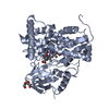
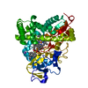
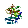
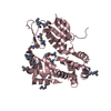
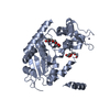
- PDB-4yru: Crystal structure of C-terminally truncated Neuronal Calcium Sens... -
+ Open data

- Basic information
| Entry | Database: PDB / ID: 4yru | ||||||
|---|---|---|---|---|---|---|---|
| Title | Crystal structure of C-terminally truncated Neuronal Calcium Sensor (NCS-1) from Rattus norvegicus | ||||||
 Components Components | Neuronal calcium sensor 1 | ||||||
 Keywords Keywords | SIGNALING PROTEIN / EF-hand / Calcium sensor / C-terminal truncation | ||||||
| Function / homology |  Function and homology information Function and homology informationcalcium-dependent protein kinase inhibitor activity / calcium sensitive guanylate cyclase activator activity / regulation of presynaptic cytosolic calcium ion concentration / presynaptic cytosol / postsynaptic cytosol / regulation of neuron projection development / regulation of synaptic vesicle exocytosis / positive regulation of exocytosis / calyx of Held / voltage-gated calcium channel activity ...calcium-dependent protein kinase inhibitor activity / calcium sensitive guanylate cyclase activator activity / regulation of presynaptic cytosolic calcium ion concentration / presynaptic cytosol / postsynaptic cytosol / regulation of neuron projection development / regulation of synaptic vesicle exocytosis / positive regulation of exocytosis / calyx of Held / voltage-gated calcium channel activity / positive regulation of calcium-mediated signaling / postsynapse / postsynaptic density / axon / glutamatergic synapse / dendrite / calcium ion binding / protein kinase binding / perinuclear region of cytoplasm / Golgi apparatus / magnesium ion binding / plasma membrane / cytoplasmSimilarity search - Function | ||||||
| Biological species |  Rattus norvegicus (Norway rat) Rattus norvegicus (Norway rat) | ||||||
| Method | X-RAY DIFFRACTION / SYNCHROTRON / MOLECULAR REPLACEMENT / Resolution: 2.8 Å | ||||||
 Authors Authors | Pandalaneni, S. / Karrupiah, V. / Mayans, O. / Derrick, J.P. / Lian, L.Y. | ||||||
 Citation Citation | Journal: J.Biol.Chem. / Year: 2015 Title: Neuronal Calcium Sensor-1 Binds the D2 Dopamine Receptor and G-protein-coupled Receptor Kinase 1 (GRK1) Peptides Using Different Modes of Interactions. Authors: Pandalaneni, S. / Karuppiah, V. / Saleem, M. / Haynes, L.P. / Burgoyne, R.D. / Mayans, O. / Derrick, J.P. / Lian, L.Y. | ||||||
| History |
|
- Structure visualization
| Structure viewer | Molecule: MolmilJmol/JSmol |
|---|
- Downloads & links
-Download
| PDBx/mmCIF format | 4yru.cif.gz | 272.3 KB | Display | PDBx/mmCIF format |
|---|---|---|---|---|
| PDB format | pdb4yru.ent.gz | 220.2 KB | Display | PDB format |
| PDBx/mmJSON format | 4yru.json.gz | Tree view | PDBx/mmJSON format | |
| Others |  Other downloads Other downloads |
-Validation report
| Arichive directory | https://data.pdbj.org/pub/pdb/validation_reports/yr/4yruftp://data.pdbj.org/pub/pdb/validation_reports/yr/4yru | HTTPS FTP |
|---|
-Related structure data
| Related structure data |  5aeqC  5aerC 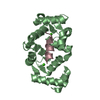 5afpC  2you S: Starting model for refinement C: citing same article ( |
|---|---|
| Similar structure data |


-Links
 PDBj
PDBj- Assembly
| Deposited unit | 
| ||||||||
|---|---|---|---|---|---|---|---|---|---|
| 1 | 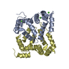
| ||||||||
| 2 | 
| ||||||||
| Unit cell |
|
-Components
| #1: Protein | / NCS-1 / Frequenin homolog / Frequenin-like protein / Frequenin-like ubiquitous protein Mass: 20543.092 Da / Num. of mol.: 4 / Fragment: UNP residues 1-177 Source method: isolated from a genetically manipulated source Source: (gene. exp.) Rattus norvegicus (Norway rat) / Gene: Ncs1, Flup, Freq / Production host:  Escherichia coli (E. coli) / References: UniProt: P62168 Escherichia coli (E. coli) / References: UniProt: P62168#2: Chemical | ChemComp-CA /   Mass: 40.078 Da / Num. of mol.: 12 / Source method: obtained synthetically / Formula: Ca Mass: 40.078 Da / Num. of mol.: 12 / Source method: obtained synthetically / Formula: Ca#3: Water | ChemComp-HOH / | Water Mass: 18.015 Da / Num. of mol.: 16 / Source method: isolated from a natural source / Formula: H2O Mass: 18.015 Da / Num. of mol.: 16 / Source method: isolated from a natural source / Formula: H2O |
|---|
-Experimental details
-Experiment
| Experiment | Method: X-RAY DIFFRACTION |
|---|
- Sample preparation
| Crystal | Density Matthews: 1.98 Å3/Da / Density % sol: 37.78 % |
|---|---|
| Crystal grow | Temperature: 293 K / Method: vapor diffusion, sitting drop / pH: 6 / Details: 0.1M MES pH6.0, 200mM NaCl,16% (w/v) PEG 6K |
-Data collection
| Diffraction | Mean temperature: 100 K |
|---|---|
| Diffraction source | Source: SYNCHROTRON / Site: Diamond  / Beamline: I02 / Wavelength: 0.97949 Å / Beamline: I02 / Wavelength: 0.97949 Å |
| Detector | Type: DECTRIS PILATUS 6M / Detector: PIXEL / Date: Jun 28, 2013 |
| Radiation | Monochromator: double crystal Si(111) / Protocol: SINGLE WAVELENGTH / Monochromatic (M) / Laue (L): M / Scattering type: x-ray |
| Radiation wavelength | Wavelength: 0.97949 Å / Relative weight: 1 |
| Reflection | Resolution: 2.8→66.59 Å / Num. obs: 16258 / % possible obs: 98 % / Redundancy: 3.2 % / Rmerge(I) obs: 0.124 / Net I/σ(I): 1.1 |
| Reflection shell | Resolution: 2.8→2.95 Å / Redundancy: 3 % / Rmerge(I) obs: 0.374 / % possible all: 98.2 |
- Processing
| Software |
| |||||||||||||||||||||||||||||||||||||||||||||||||||||||||||||||||||||||||||||||||||||||||||||||||||||||||||||||||||||||||||||
|---|---|---|---|---|---|---|---|---|---|---|---|---|---|---|---|---|---|---|---|---|---|---|---|---|---|---|---|---|---|---|---|---|---|---|---|---|---|---|---|---|---|---|---|---|---|---|---|---|---|---|---|---|---|---|---|---|---|---|---|---|---|---|---|---|---|---|---|---|---|---|---|---|---|---|---|---|---|---|---|---|---|---|---|---|---|---|---|---|---|---|---|---|---|---|---|---|---|---|---|---|---|---|---|---|---|---|---|---|---|---|---|---|---|---|---|---|---|---|---|---|---|---|---|---|---|---|
| Refinement | Method to determine structure: MOLECULAR REPLACEMENT Starting model: 2you 2you Resolution: 2.8→66.59 Å / SU ML: 0.4 / Cross valid method: FREE R-VALUE / σ(F): 1.37 / Phase error: 34.77 / Stereochemistry target values: ML
| |||||||||||||||||||||||||||||||||||||||||||||||||||||||||||||||||||||||||||||||||||||||||||||||||||||||||||||||||||||||||||||
| Solvent computation | Shrinkage radii: 0.9 Å / VDW probe radii: 1.11 Å / Solvent model: FLAT BULK SOLVENT MODEL | |||||||||||||||||||||||||||||||||||||||||||||||||||||||||||||||||||||||||||||||||||||||||||||||||||||||||||||||||||||||||||||
| Refinement step | Cycle: LAST / Resolution: 2.8→66.59 Å
| |||||||||||||||||||||||||||||||||||||||||||||||||||||||||||||||||||||||||||||||||||||||||||||||||||||||||||||||||||||||||||||
| Refine LS restraints |
| |||||||||||||||||||||||||||||||||||||||||||||||||||||||||||||||||||||||||||||||||||||||||||||||||||||||||||||||||||||||||||||
| LS refinement shell |
| |||||||||||||||||||||||||||||||||||||||||||||||||||||||||||||||||||||||||||||||||||||||||||||||||||||||||||||||||||||||||||||
| Refinement TLS params. | Method: refined / Refine-ID: X-RAY DIFFRACTION
| |||||||||||||||||||||||||||||||||||||||||||||||||||||||||||||||||||||||||||||||||||||||||||||||||||||||||||||||||||||||||||||
| Refinement TLS group |
|
