 Movie
Movie Controller

[English] 日本語
 Yorodumi
- PDB-4ync: OYE1 W116A COMPLEXED WITH (Z)-METHYL-3-CYANO-3-PHENYLACRYLATE IN ... -
+ Open data

- Basic information
| Entry | Database: PDB / ID: 4ync | |||||||||
|---|---|---|---|---|---|---|---|---|---|---|
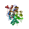
| Title | OYE1 W116A COMPLEXED WITH (Z)-METHYL-3-CYANO-3-PHENYLACRYLATE IN A NON PRODUCTIVE BINDING MODE | |||||||||
 Components Components | NADPH dehydrogenase 1 | |||||||||
 Keywords Keywords | OXIDOREDUCTASE / CATALYTIC ACTIVITY / FMN BINDING / OLD YELLOW ENZYME | |||||||||
| Function / homology |  Function and homology informationNADPH dehydrogenase / pentaerythritol trinitrate reductase activity / NADPH dehydrogenase activity / FMN binding Function and homology informationNADPH dehydrogenase / pentaerythritol trinitrate reductase activity / NADPH dehydrogenase activity / FMN bindingSimilarity search - Function | |||||||||
| Biological species |  Saccharomyces pastorianus (lager yeast) Saccharomyces pastorianus (lager yeast) | |||||||||
| Method | X-RAY DIFFRACTION / SYNCHROTRON / MOLECULAR REPLACEMENT / Resolution: 1.498 Å | |||||||||
 Authors Authors | Santangelo, S. / Brenna, E. / Stewart, J.D. / Powell III, R.W. | |||||||||
 Citation Citation | Journal: Adv.Synth.Catal. / Year: 2015 Title: Opposite Enantioselectivity in the Bioreduction of (Z)-beta-Aryl-beta-cyanoacrylates Mediated by the Tryptophan 116 Mutants of Old Yellow Enzyme 1: Synthetic Approach to (R)- and (S)-beta-Aryl-gamma-lactams Authors: Brenna, E. / Crotti, M. / Gatti, F.G. / Monti, D. / Parmeggiani, F. / Powell III, R.W. / Santangelo, S. / Stewart, J.D. | |||||||||
| History |
|
- Structure visualization
| Structure viewer | Molecule: MolmilJmol/JSmol |
|---|
- Downloads & links
-Download
| PDBx/mmCIF format | 4ync.cif.gz | 109.9 KB | Display | PDBx/mmCIF format |
|---|---|---|---|---|
| PDB format | pdb4ync.ent.gz | 81.7 KB | Display | PDB format |
| PDBx/mmJSON format | 4ync.json.gz | Tree view | PDBx/mmJSON format | |
| Others |  Other downloads Other downloads |
-Validation report
| Arichive directory | https://data.pdbj.org/pub/pdb/validation_reports/yn/4yncftp://data.pdbj.org/pub/pdb/validation_reports/yn/4ync | HTTPS FTP |
|---|
-Related structure data
| Related structure data |  4yilC  4gbuS S: Starting model for refinement C: citing same article ( |
|---|---|
| Similar structure data |


-Links
 PDBj
PDBj- Assembly
| Deposited unit | 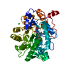
| |||||||||
|---|---|---|---|---|---|---|---|---|---|---|
| 1 |
| |||||||||
| Unit cell |
| |||||||||
| Components on special symmetry positions |
|
-Components
| #1: Protein | / Old yellow enzyme 1 Mass: 44566.953 Da / Num. of mol.: 1 / Mutation: W116A Source method: isolated from a genetically manipulated source Source: (gene. exp.) Saccharomyces pastorianus (lager yeast)Gene: OYE1 / Plasmid: pET3b / Details (production host): pSA01 / Production host:  Escherichia coli BL21(DE3) (bacteria) / References: UniProt: Q02899, NADPH dehydrogenase Escherichia coli BL21(DE3) (bacteria) / References: UniProt: Q02899, NADPH dehydrogenase |
|---|---|
| #2: Chemical | ChemComp-FMN / Flavin mononucleotide  Mass: 456.344 Da / Num. of mol.: 1 / Source method: obtained synthetically / Formula: C17H21N4O9P Mass: 456.344 Da / Num. of mol.: 1 / Source method: obtained synthetically / Formula: C17H21N4O9P |
| #3: Chemical | ChemComp-4EG /   Mass: 187.195 Da / Num. of mol.: 1 / Source method: obtained synthetically / Formula: C11H9NO2 Mass: 187.195 Da / Num. of mol.: 1 / Source method: obtained synthetically / Formula: C11H9NO2 |
| #4: Chemical | ChemComp-CL / Chloride  Mass: 35.453 Da / Num. of mol.: 1 / Source method: obtained synthetically / Formula: Cl Mass: 35.453 Da / Num. of mol.: 1 / Source method: obtained synthetically / Formula: Cl |
| #5: Water | ChemComp-HOH / Water Mass: 18.015 Da / Num. of mol.: 545 / Source method: isolated from a natural source / Formula: H2O Mass: 18.015 Da / Num. of mol.: 545 / Source method: isolated from a natural source / Formula: H2O |
-Experimental details
-Experiment
| Experiment | Method: X-RAY DIFFRACTION / Number of used crystals: 1 |
|---|
- Sample preparation
| Crystal | Density Matthews: 2.41 Å3/Da / Density % sol: 48.91 % |
|---|---|
| Crystal grow | Temperature: 277 K / Method: vapor diffusion, hanging drop / pH: 8.3 / Details: 0.2M MgCl2, 0.1M NaHEPES, 35% PEG 400 |
-Data collection
| Diffraction | Mean temperature: 100 K |
|---|---|
| Diffraction source | Source: SYNCHROTRON / Site: NSLS  / Beamline: X6A / Wavelength: 0.95 Å / Beamline: X6A / Wavelength: 0.95 Å |
| Detector | Type: ADSC QUANTUM 270 / Detector: CCD / Date: Aug 30, 2014 |
| Radiation | Protocol: SINGLE WAVELENGTH / Monochromatic (M) / Laue (L): M / Scattering type: x-ray |
| Radiation wavelength | Wavelength: 0.95 Å / Relative weight: 1 |
| Reflection | Resolution: 1.498→27.75 Å / Num. all: 70253 / Num. obs: 70253 / % possible obs: 99.76 % / Redundancy: 14.7 % / Rmerge(I) obs: 0.087 / Net I/σ(I): 29.05 |
| Reflection shell | Resolution: 1.498→1.551 Å / Redundancy: 13.3 % / Rmerge(I) obs: 0.777 / Mean I/σ(I) obs: 4.65 / % possible all: 99.39 |
- Processing
| Software |
| |||||||||||||||||||||||||||||||||||||||||||||||||||||||||||||||||||||||||||||||||||||||||||||||||||||||||
|---|---|---|---|---|---|---|---|---|---|---|---|---|---|---|---|---|---|---|---|---|---|---|---|---|---|---|---|---|---|---|---|---|---|---|---|---|---|---|---|---|---|---|---|---|---|---|---|---|---|---|---|---|---|---|---|---|---|---|---|---|---|---|---|---|---|---|---|---|---|---|---|---|---|---|---|---|---|---|---|---|---|---|---|---|---|---|---|---|---|---|---|---|---|---|---|---|---|---|---|---|---|---|---|---|---|---|
| Refinement | Method to determine structure: MOLECULAR REPLACEMENT Starting model: 4GBU Resolution: 1.498→27.751 Å / SU ML: 0.12 / Cross valid method: FREE R-VALUE / σ(F): 1.34 / Phase error: 16.33 / Stereochemistry target values: ML
| |||||||||||||||||||||||||||||||||||||||||||||||||||||||||||||||||||||||||||||||||||||||||||||||||||||||||
| Solvent computation | Shrinkage radii: 0.9 Å / VDW probe radii: 1.11 Å / Solvent model: FLAT BULK SOLVENT MODEL | |||||||||||||||||||||||||||||||||||||||||||||||||||||||||||||||||||||||||||||||||||||||||||||||||||||||||
| Displacement parameters | Biso max: 79.04 Å2 / Biso mean: 18.988 Å2 / Biso min: 9.97 Å2 | |||||||||||||||||||||||||||||||||||||||||||||||||||||||||||||||||||||||||||||||||||||||||||||||||||||||||
| Refinement step | Cycle: final / Resolution: 1.498→27.751 Å
| |||||||||||||||||||||||||||||||||||||||||||||||||||||||||||||||||||||||||||||||||||||||||||||||||||||||||
| Refine LS restraints |
| |||||||||||||||||||||||||||||||||||||||||||||||||||||||||||||||||||||||||||||||||||||||||||||||||||||||||
| LS refinement shell | Refine-ID: X-RAY DIFFRACTION / Total num. of bins used: 14
|
