 Movie
Movie Controller

[English] 日本語
 Yorodumi
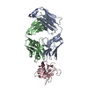
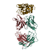
- PDB-4ybl: Crystal structure of the stabilized inner domain of clade A/E HIV... -
+ Open data

- Basic information
| Entry | Database: PDB / ID: 4ybl | ||||||
|---|---|---|---|---|---|---|---|
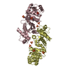
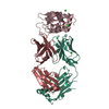
| Title | Crystal structure of the stabilized inner domain of clade A/E HIV-1 gp120 in complex with the ADCC mediating ANTI-HIV-1 antibody A32 | ||||||
 Components Components |
| ||||||
 Keywords Keywords |  VIRAL PROTEIN/IMMUNE SYSTEM / ADCC / NON-NEUTRALIZING / ANTI-HIV-1 ENV ANTIBODY A32 / CD4I ANTIBODY / VIRAL GLYCOPROTEIN GP120 / HIV-1 ENV / VIRAL PROTEIN-IMMUNE SYSTEM complex VIRAL PROTEIN/IMMUNE SYSTEM / ADCC / NON-NEUTRALIZING / ANTI-HIV-1 ENV ANTIBODY A32 / CD4I ANTIBODY / VIRAL GLYCOPROTEIN GP120 / HIV-1 ENV / VIRAL PROTEIN-IMMUNE SYSTEM complex | ||||||
| Function / homology | Gp120 core superfamily / Envelope glycoprotein GP120 / Human immunodeficiency virus 1, envelope glycoprotein Gp120 / viral envelope / Immunoglobulins / Immunoglobulin-like / Sandwich / Mainly Beta / clade A/E 93TH057 HIV-1 gp120 core Function and homology information Function and homology information | ||||||
| Biological species |   Human immunodeficiency virus 1 Human immunodeficiency virus 1 Homo sapiens (human) Homo sapiens (human) | ||||||
| Method | X-RAY DIFFRACTION / SYNCHROTRON / MOLECULAR REPLACEMENT / Resolution: 3.1 Å | ||||||
 Authors Authors | Tolbert, W.D. / Gohain, N. / Pazgier, M. | ||||||
| Funding support |  United States, 1items United States, 1items
| ||||||
 Citation Citation | Journal: Structure / Year: 2016 Title: Paring Down HIV Env: Design and Crystal Structure of a Stabilized Inner Domain of HIV-1 gp120 Displaying a Major ADCC Target of the A32 Region. Authors: Tolbert, W.D. / Gohain, N. / Veillette, M. / Chapleau, J.P. / Orlandi, C. / Visciano, M.L. / Ebadi, M. / DeVico, A.L. / Fouts, T.R. / Finzi, A. / Lewis, G.K. / Pazgier, M. | ||||||
| History |
|
- Structure visualization
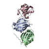
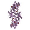
| Structure viewer | Molecule: MolmilJmol/JSmol |
|---|
- Downloads & links
-Download
| PDBx/mmCIF format | 4ybl.cif.gz | 437.9 KB | Display | PDBx/mmCIF format |
|---|---|---|---|---|
| PDB format | pdb4ybl.ent.gz | 364.8 KB | Display | PDB format |
| PDBx/mmJSON format | 4ybl.json.gz | Tree view | PDBx/mmJSON format | |
| Others |  Other downloads Other downloads |
-Validation report
| Arichive directory | https://data.pdbj.org/pub/pdb/validation_reports/yb/4yblftp://data.pdbj.org/pub/pdb/validation_reports/yb/4ybl | HTTPS FTP |
|---|
-Related structure data
| Related structure data | 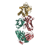 4yc2C 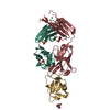 5fcuC 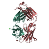 3tnmS  4rqh S: Starting model for refinement C: citing same article ( |
|---|---|
| Similar structure data |


-Links
 PDBj
PDBj


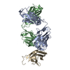
- Assembly
| Deposited unit | 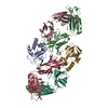
| ||||||||
|---|---|---|---|---|---|---|---|---|---|
| 1 | 
| ||||||||
| 2 | 
| ||||||||
| Unit cell |
| ||||||||
| Details | The complex was purified by gel filtration prior to crystallization. |
-Components
| #1: Protein | Mass: 17236.402 Da / Num. of mol.: 2 / Mutation: V65C, S115C Source method: isolated from a genetically manipulated source Source: (gene. exp.) Human immunodeficiency virus 1 / Strain: clade A/E / Cell line (production host): HEK GnT1- 293 cells / Production host: Homo sapiens (human) / References: UniProt: A0A0M3KKW9*PLUS#2: Antibody | Mass: 23991.855 Da / Num. of mol.: 2 Source method: isolated from a genetically manipulated source Source: (gene. exp.) Homo sapiens (human) / Cell line (production host): HEK 293 cells / Production host: Homo sapiens (human)#3: Antibody | Mass: 22193.434 Da / Num. of mol.: 2 Source method: isolated from a genetically manipulated source Source: (gene. exp.) Homo sapiens (human) / Cell line (production host): HEK 293 cells / Production host: Homo sapiens (human)Sequence details | The sequence of the clade A/E gp120 is based on the HIV-1 clade A/E gp120 sequence in PDB ID 3TGT. ...The sequence of the clade A/E gp120 is based on the HIV-1 clade A/E gp120 sequence in PDB ID 3TGT. The sequence was engineered to remove the outer domain of gp120 and consists of the N-terminal sequence plus some of the C-terminal sequence. | |
|---|
-Experimental details
-Experiment
| Experiment | Method: X-RAY DIFFRACTION |
|---|
- Sample preparation
| Crystal |
| ||||||||||||
|---|---|---|---|---|---|---|---|---|---|---|---|---|---|
| Crystal grow | Temperature: 295 K / Method: vapor diffusion, hanging drop / pH: 8.5 / Details: 18-22% PEG 6000 or 8000 0.1 M Tris-HCl pH 8.5 |
-Data collection
| Diffraction |
| ||||||||||||||||||
|---|---|---|---|---|---|---|---|---|---|---|---|---|---|---|---|---|---|---|---|
| Diffraction source |
| ||||||||||||||||||
| Detector |
| ||||||||||||||||||
| Radiation |
| ||||||||||||||||||
| Radiation wavelength |
| ||||||||||||||||||
| Reflection | Resolution: 3.1→50 Å / Num. all: 21415 / Num. obs: 19959 / % possible obs: 93.2 % / Observed criterion σ(F): 0 / Redundancy: 8.3 % / Rmerge(I) obs: 0.186 / Net I/σ(I): 14.6 | ||||||||||||||||||
| Reflection shell | Resolution: 3.1→3.15 Å / Redundancy: 8.3 % / Rmerge(I) obs: 0.955 / Mean I/σ(I) obs: 1.3 / % possible all: 96.8 |
- Processing
| Software |
| ||||||||||||||||||||||||||||||||||||||||||||||||||||||||||||||||||||||||||||||||||||||||||||||||||||||||||||||||||||||||||||||||||||||||||||||||||||||||||||||||||||||||||||||||||||||
|---|---|---|---|---|---|---|---|---|---|---|---|---|---|---|---|---|---|---|---|---|---|---|---|---|---|---|---|---|---|---|---|---|---|---|---|---|---|---|---|---|---|---|---|---|---|---|---|---|---|---|---|---|---|---|---|---|---|---|---|---|---|---|---|---|---|---|---|---|---|---|---|---|---|---|---|---|---|---|---|---|---|---|---|---|---|---|---|---|---|---|---|---|---|---|---|---|---|---|---|---|---|---|---|---|---|---|---|---|---|---|---|---|---|---|---|---|---|---|---|---|---|---|---|---|---|---|---|---|---|---|---|---|---|---|---|---|---|---|---|---|---|---|---|---|---|---|---|---|---|---|---|---|---|---|---|---|---|---|---|---|---|---|---|---|---|---|---|---|---|---|---|---|---|---|---|---|---|---|---|---|---|---|---|
| Refinement | Method to determine structure: MOLECULAR REPLACEMENT Starting model: 3TNM and 4RQH Resolution: 3.1→50 Å / Cor.coef. Fo:Fc: 0.924 / Cor.coef. Fo:Fc free: 0.877 / SU B: 74.752 / SU ML: 0.551 / Cross valid method: THROUGHOUT / ESU R Free: 0.629 / Stereochemistry target values: MAXIMUM LIKELIHOOD / Details: HYDROGENS HAVE BEEN ADDED IN THE RIDING POSITIONS
| ||||||||||||||||||||||||||||||||||||||||||||||||||||||||||||||||||||||||||||||||||||||||||||||||||||||||||||||||||||||||||||||||||||||||||||||||||||||||||||||||||||||||||||||||||||||
| Solvent computation | Ion probe radii: 0.8 Å / Shrinkage radii: 0.8 Å / VDW probe radii: 1.2 Å / Solvent model: MASK | ||||||||||||||||||||||||||||||||||||||||||||||||||||||||||||||||||||||||||||||||||||||||||||||||||||||||||||||||||||||||||||||||||||||||||||||||||||||||||||||||||||||||||||||||||||||
| Displacement parameters | Biso mean: 94.206 Å2
| ||||||||||||||||||||||||||||||||||||||||||||||||||||||||||||||||||||||||||||||||||||||||||||||||||||||||||||||||||||||||||||||||||||||||||||||||||||||||||||||||||||||||||||||||||||||
| Refinement step | Cycle: 1 / Resolution: 3.1→50 Å
| ||||||||||||||||||||||||||||||||||||||||||||||||||||||||||||||||||||||||||||||||||||||||||||||||||||||||||||||||||||||||||||||||||||||||||||||||||||||||||||||||||||||||||||||||||||||
| Refine LS restraints |
|