 Movie
Movie Controller

[English] 日本語
 Yorodumi
- PDB-5eii: Structural determination of an protein complex of a Fab with incr... -
+ Open data

- Basic information
| Entry | Database: PDB / ID: 5eii | ||||||||||||
|---|---|---|---|---|---|---|---|---|---|---|---|---|---|
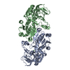
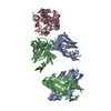
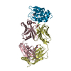
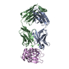
| Title | Structural determination of an protein complex of a Fab with increased solubility | ||||||||||||
 Components Components |
| ||||||||||||
 Keywords Keywords |  IMMUNE SYSTEM / Antibody / Fab / Asf1 / Structural Genomics / PSI-Biology / Chaperone-Enabled Studies of Epigenetic Regulation Enzymes / CEBS IMMUNE SYSTEM / Antibody / Fab / Asf1 / Structural Genomics / PSI-Biology / Chaperone-Enabled Studies of Epigenetic Regulation Enzymes / CEBS | ||||||||||||
| Function / homology |  Function and homology information: / Formation of Senescence-Associated Heterochromatin Foci (SAHF) / H3 histone acetyltransferase complex / acetyltransferase activator activity / DNA replication-dependent chromatin assembly / nucleosome disassembly / : / silent mating-type cassette heterochromatin formation / subtelomeric heterochromatin formation / negative regulation of DNA damage checkpoint ...: / Formation of Senescence-Associated Heterochromatin Foci (SAHF) / H3 histone acetyltransferase complex / acetyltransferase activator activity / DNA replication-dependent chromatin assembly / nucleosome disassembly / : / silent mating-type cassette heterochromatin formation / subtelomeric heterochromatin formation / negative regulation of DNA damage checkpoint / regulation of DNA repair / positive regulation of transcription elongation by RNA polymerase II / regulation of protein phosphorylation / nucleosome assembly / chromatin organization / histone binding / regulation of gene expression / chromosome, telomeric region / regulation of transcription by RNA polymerase II / nucleus / cytosol Function and homology information: / Formation of Senescence-Associated Heterochromatin Foci (SAHF) / H3 histone acetyltransferase complex / acetyltransferase activator activity / DNA replication-dependent chromatin assembly / nucleosome disassembly / : / silent mating-type cassette heterochromatin formation / subtelomeric heterochromatin formation / negative regulation of DNA damage checkpoint ...: / Formation of Senescence-Associated Heterochromatin Foci (SAHF) / H3 histone acetyltransferase complex / acetyltransferase activator activity / DNA replication-dependent chromatin assembly / nucleosome disassembly / : / silent mating-type cassette heterochromatin formation / subtelomeric heterochromatin formation / negative regulation of DNA damage checkpoint / regulation of DNA repair / positive regulation of transcription elongation by RNA polymerase II / regulation of protein phosphorylation / nucleosome assembly / chromatin organization / histone binding / regulation of gene expression / chromosome, telomeric region / regulation of transcription by RNA polymerase II / nucleus / cytosolSimilarity search - Function | ||||||||||||
| Biological species |  Homo sapiens (human) Homo sapiens (human) Saccharomyces cerevisiae (brewer's yeast) Saccharomyces cerevisiae (brewer's yeast) | ||||||||||||
| Method | X-RAY DIFFRACTION / SYNCHROTRON / Resolution: 2.44 Å | ||||||||||||
 Authors Authors | Bailey, L.J. / Kossiakoff, A.A. / Chaperone-Enabled Studies of Epigenetic Regulation Enzymes (CEBS) | ||||||||||||
| Funding support |  United States, 3items United States, 3items
| ||||||||||||
 Citation Citation | Journal: To Be Published Title: Structural determination of an protein complex of a Fab with increased solubility Authors: Bailey, L.J. / Kossiakoff, A.A. / Schaefer, Z.P. #1: Journal: To Be PublishedTitle: A polar ring endows improved specificity to an antibody fragment Authors: Schaefer, Z.P. / Bailey, L.J. / Kossiakoff, A.A. | ||||||||||||
| History |
|
- Structure visualization
| Structure viewer | Molecule: MolmilJmol/JSmol |
|---|
- Downloads & links
-Download
| PDBx/mmCIF format | 5eii.cif.gz | 451.1 KB | Display | PDBx/mmCIF format |
|---|---|---|---|---|
| PDB format | pdb5eii.ent.gz | 381.8 KB | Display | PDB format |
| PDBx/mmJSON format | 5eii.json.gz | Tree view | PDBx/mmJSON format | |
| Others |  Other downloads Other downloads |
-Validation report
| Arichive directory | https://data.pdbj.org/pub/pdb/validation_reports/ei/5eiiftp://data.pdbj.org/pub/pdb/validation_reports/ei/5eii | HTTPS FTP |
|---|
-Related structure data
| Similar structure data |
|---|

-Links
 PDBj
PDBj

- Assembly
| Deposited unit | 
| ||||||||
|---|---|---|---|---|---|---|---|---|---|
| 1 | 
| ||||||||
| 2 | 
| ||||||||
| Unit cell |
|
-Components
| #1: Antibody | Fragment antigen-binding Mass: 25084.887 Da / Num. of mol.: 2 Source method: isolated from a genetically manipulated source Source: (gene. exp.) Homo sapiens (human) / Production host:  Escherichia coli (E. coli) Escherichia coli (E. coli)#2: Antibody | Fragment antigen-bindingMass: 23298.760 Da / Num. of mol.: 2 Source method: isolated from a genetically manipulated source Source: (gene. exp.) Homo sapiens (human) / Production host: Escherichia coli (E. coli)#3: Protein | Mass: 17746.012 Da / Num. of mol.: 2 / Fragment: UNP residues 1-156 Source method: isolated from a genetically manipulated source Source: (gene. exp.) Saccharomyces cerevisiae (brewer's yeast)Gene: ASF1, CIA1, YJL115W, J0755 / Production host: Escherichia coli (E. coli) / References: UniProt: P32447#4: Water | ChemComp-HOH / | Water Mass: 18.015 Da / Num. of mol.: 429 / Source method: isolated from a natural source / Formula: H2O Mass: 18.015 Da / Num. of mol.: 429 / Source method: isolated from a natural source / Formula: H2O |
|---|
-Experimental details
-Experiment
| Experiment | Method: X-RAY DIFFRACTION / Number of used crystals: 1 |
|---|
- Sample preparation
| Crystal | Density Matthews: 3.13 Å3/Da / Density % sol: 60.71 % |
|---|---|
| Crystal grow | Temperature: 293 K / Method: liquid diffusion Details: Crystals were grown by hanging drop liquid diffusion by adding a solution containing 20% PEG 3350 and Sodium Acetate, pH 5.5 |
-Data collection
| Diffraction | Mean temperature: 80 K |
|---|---|
| Diffraction source | Source: SYNCHROTRON / Site: APS / Beamline: 23-ID-B / Wavelength: 0.987 Å |
| Detector | Type: MAR scanner 300 mm plate / Detector: IMAGE PLATE / Date: Dec 13, 2013 |
| Radiation | Protocol: SINGLE WAVELENGTH / Monochromatic (M) / Laue (L): M / Scattering type: x-ray |
| Radiation wavelength | Wavelength: 0.987 Å / Relative weight: 1 |
| Reflection | Resolution: 2.43723→46.3844 Å / Num. obs: 61283 / % possible obs: 99.4499 % / Redundancy: 3.6 % / Net I/σ(I): 12.1 |
- Processing
| Software |
| |||||||||||||||||||||||||||||||||||||||||||||||||||||||||||||||||||||||||||||||||||||||||||||||||||||||||||||||||||||||||||||||||||||||||||||||||||||||||||||||||
|---|---|---|---|---|---|---|---|---|---|---|---|---|---|---|---|---|---|---|---|---|---|---|---|---|---|---|---|---|---|---|---|---|---|---|---|---|---|---|---|---|---|---|---|---|---|---|---|---|---|---|---|---|---|---|---|---|---|---|---|---|---|---|---|---|---|---|---|---|---|---|---|---|---|---|---|---|---|---|---|---|---|---|---|---|---|---|---|---|---|---|---|---|---|---|---|---|---|---|---|---|---|---|---|---|---|---|---|---|---|---|---|---|---|---|---|---|---|---|---|---|---|---|---|---|---|---|---|---|---|---|---|---|---|---|---|---|---|---|---|---|---|---|---|---|---|---|---|---|---|---|---|---|---|---|---|---|---|---|---|---|---|---|
| Refinement | Resolution: 2.44→46.384 Å / SU ML: 0.34 / Cross valid method: FREE R-VALUE / σ(F): 1.35 / Phase error: 25.01 / Stereochemistry target values: ML
| |||||||||||||||||||||||||||||||||||||||||||||||||||||||||||||||||||||||||||||||||||||||||||||||||||||||||||||||||||||||||||||||||||||||||||||||||||||||||||||||||
| Solvent computation | Shrinkage radii: 0.9 Å / VDW probe radii: 1.11 Å / Solvent model: FLAT BULK SOLVENT MODEL | |||||||||||||||||||||||||||||||||||||||||||||||||||||||||||||||||||||||||||||||||||||||||||||||||||||||||||||||||||||||||||||||||||||||||||||||||||||||||||||||||
| Refinement step | Cycle: LAST / Resolution: 2.44→46.384 Å
| |||||||||||||||||||||||||||||||||||||||||||||||||||||||||||||||||||||||||||||||||||||||||||||||||||||||||||||||||||||||||||||||||||||||||||||||||||||||||||||||||
| Refine LS restraints |
| |||||||||||||||||||||||||||||||||||||||||||||||||||||||||||||||||||||||||||||||||||||||||||||||||||||||||||||||||||||||||||||||||||||||||||||||||||||||||||||||||
| LS refinement shell |
| |||||||||||||||||||||||||||||||||||||||||||||||||||||||||||||||||||||||||||||||||||||||||||||||||||||||||||||||||||||||||||||||||||||||||||||||||||||||||||||||||
| Refinement TLS params. | Method: refined / Origin x: 6.3942 Å / Origin y: 5.5826 Å / Origin z: 34.589 Å
| |||||||||||||||||||||||||||||||||||||||||||||||||||||||||||||||||||||||||||||||||||||||||||||||||||||||||||||||||||||||||||||||||||||||||||||||||||||||||||||||||
| Refinement TLS group | Selection details: all |
