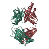
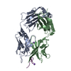
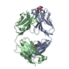
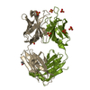
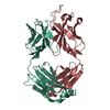
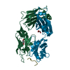
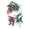
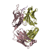
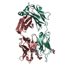
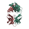
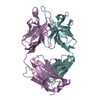
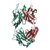
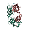
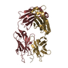
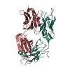
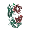
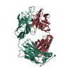
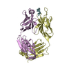
Entry Database : PDB / ID : 4xxdTitle Crystal Structure of mid-region amyloid beta capture by solanezumab Amyloid-beta fragment Fab Heavy Chain Fab Light Chain Keywords / / Function / homology Function Domain/homology Component
/ / / / / / / / / / / / / / / / / / / / / / / / / / / / / / / / / / / / / / / / / / / / / / / / / / / / / / / / / / / / / / / / / / / / / / / / / / / / / / / / / / / / / / / / / / / / / / / / / / / / / / / / / / / / / / / / / / / / / / / / / / / / / / / / / / / / / / / / / / / / / / / / / / / / / / / / / / / / / / / / Biological species Homo sapiens (human)Method / / / Resolution : 2.41 Å Authors Hermans, S.J. / Crespi, G.A.N. / Parker, M.W. / Miles, L.A. Funding support Organization Grant number Country National Health and Medical Council Project Grant APP1021935
Journal : Sci Rep / Year : 2015Title : Molecular basis for mid-region amyloid-beta capture by leading Alzheimer's disease immunotherapies.Authors : Crespi, G.A. / Hermans, S.J. / Parker, M.W. / Miles, L.A. History Deposition Jan 30, 2015 Deposition site / Processing site Revision 1.0 Apr 29, 2015 Provider / Type Revision 1.1 Jul 29, 2015 Group / Structure summaryRevision 1.2 Aug 23, 2017 Group / Derived calculations / Source and taxonomyCategory diffrn_detector / diffrn_source ... diffrn_detector / diffrn_source / entity_src_gen / pdbx_entity_src_syn / pdbx_struct_oper_list Item _diffrn_detector.detector / _diffrn_source.pdbx_synchrotron_site ... _diffrn_detector.detector / _diffrn_source.pdbx_synchrotron_site / _entity_src_gen.pdbx_alt_source_flag / _pdbx_entity_src_syn.pdbx_alt_source_flag / _pdbx_struct_oper_list.symmetry_operation Revision 1.3 Sep 27, 2023 Group / Database references / Refinement descriptionCategory chem_comp_atom / chem_comp_bond ... chem_comp_atom / chem_comp_bond / database_2 / pdbx_initial_refinement_model Item / _database_2.pdbx_database_accession
Show all Show less
 Movie
Movie Controller

 Yorodumi
 Open data
 Basic information
 Components
 Keywords
 IMMUNE SYSTEM /
  Function and homology information
 Authors
 Australia, 1items
  Citation
 Structure visualization
 Downloads & links
 Other downloads
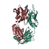


 PDBj
PDBj


 Assembly
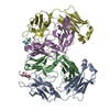

 Mass: 18.015 Da / Num. of mol.: 278 / Source method: isolated from a natural source / Formula: H2O
 Sample preparation
 Processing